Picturing Science and Engineering Felice C. Frankel MIT Press (2018)
Researchers: Felice Frankel will make photographers of you all. Photography — both macro- and microscopic — is an essential tool in science, because pictures are both intuitive and emotionally resonant. They provide insight in an instant. If you lack the skills to take exceptional photos that might make it onto a journal cover or go viral on Twitter, Frankel, a seasoned scientific photographer, hands you the keys in her latest book, Picturing Science and Engineering.
Hers is a practical manual featuring simple techniques and easily accessible tools, such as flatbed scanners and smartphones, as well as advice on lighting, composition and resolution. Frankel also covers the nuances of image manipulation appropriate to a scientific context. Massive and beautifully illustrated, the book is a trove of clear and concise recipes in granular detail. Frankel exhorts the researcher to “learn to see” through practice and a sense of play. That process can advance researchers’ own knowledge, along with the ultimate good — public understanding. There is an imperative here: document well, early, and often, so we can share in the discovery.

Credit: Felice Frankel
The joys of a flatbed scanner. This inexpensive tool can create amazing scientific pictures, such as these images of the sea sponge Euplectella aspergillum in a dry state. The top image is in very high resolution, allowing for the stunning close-up below. Zoomed in, we can clearly see silica fibres around 50 micrometres wide. A piece of black velvet serves as the background.

Credit: Felice Frankel
Simple experimentation. Frankel encourages creative play and trial and error. Here are two images of the same set-up of a row of devices on a flatbed scanner, with very different results. The difference was achieved simply by closing (left) and opening (right) the cover of the scanner. A good scanner will have options for both transmitted light (coming from above, in the cover) and reflected light (from below).

Credit: Felice Frankel
Composition. Here are two Petri dishes of the bacterium Escherichia coli, placed on a light box and photographed with a camera. The radial pattern of the bacteria is eye-catching, but what makes this stand out is the careful choreography of raw materials (the patterns in each dish) and the idea of showing just part of each dish. So think seriously about composition. Why photograph a single object if there are two or three? Why create an image with one object perfectly centred? Document your work as if it were a piece of fine art, rather than a figure in a patent application.

Credit: Felice Frankel
Using a camera correctly. Despite increasing technological sophistication, the basics of cameras remain the same. Frankel walks the reader through core concepts in photography, such as the relationship between aperture and depth of field. As the images here show, the size of your aperture determines how much of your image will be in focus, and how much light is let into the camera. So if you narrow your aperture to achieve greater focus on both the foreground and background, you need to adjust the amount of time the shutter is open — and think about adding a tripod to avoid blurriness.

Credit: Felice Frankel
Lighting is everything. The key is in the name: photo-graphy, drawing with light. Light itself is the medium. Frankel offers practical suggestions, such as using different sources of light (daylight, incandescent bulbs, ultraviolet lamps), locations (a variety of heights and angles) and techniques (mirrors, reflective ‘bounce cards’, torches). Creative lighting will enhance your experimental set-up to make it more relevant and engaging, as was done with this chamber that produces silicon wafers (above, right). Frankel also recommends using a light box or scanner to illuminate objects to be photographed with a camera on a tripod.

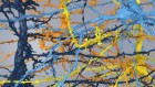
Fluorescence. Capturing fluorescent materials requires UV light and long exposures, which means the shutter is open for a long time. This kind of work demands a tripod. For this image of cadmium selenide nanocrystals (quantum dots), the exposure was about 4 seconds, captured using two UV lamps. You can also combine UV light with the ambient light in a room.

Credit: Felice Frankel
Microscopy. Frankel frames her use of microscopes as explanatory rather than exploratory: she is interested in using these powerful tools to make highly aesthetic images that illuminate the science. She has experimented with different kinds of microscopy, as seen in this range of views of a piece of old computer memory core, taken (left to right) with a stereo microscope, a compound microscope and a scanning electron microscope (false colour). Frankel encourages readers to try photographing the same material in different ways, with different tools, as a route to discovery.

Credit: Felice Frankel
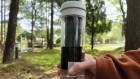
Phone cameras. Most of us have experienced how the cameras in our phones have rapidly improved: it’s now possible to take publishable images with them. In terms of resolution, they still do not compete with flatbed scanners and SLR cameras, but they are good for early concept work, such as creating a series of draft images that inform a later shoot with a camera. Phone cameras can also be used with microscopes, aided by tools such as the one pictured (left), which can position your phone over the eyepiece. The image on the right, of water bubbles in diluted coffee, was created in this way.
 Mapping the topical space
 Data visualization: Drawing out the meaning
 Picturing science