
Key Points
-
Correct diagnosis and treatment planning is crucial.
-
A diagnostic wax up is essential to allow conservative preparation and prototype fabrication and thus verifying any change in the occlusal scheme.
-
Porcelain laminate veneers can be a conservative treatment modality for treating signs and symptoms of occlusal disease.
-
A thorough understanding of occlusion is essential for any rehabilitation case be it functional or aesthetic.
Key Points
Aesthetics and cosmetics
-
1
Aesthetic changes with four anterior units
-
2
Smile lifts - a functional and aesthetic perspective
-
3
Increasing occlusal vertical dimension - why, when and how
Abstract
Cosmetic dentistry has evolved with the advent of more robust porcelain materials and ever-stronger bonding agents. This series of three articles aims to provide a practical overview of what is now possible both functionally and cosmetically from the preparation of a small number of teeth, through a whole smile, to full mouth rehabilitation. A complete diagnosis is the starting point to planning any cosmetic or functional changes. Guidance is given on the techniques used but adequate training must be considered essential before embarking upon modification in occlusal schemes or even minor adjustments in smile design. Porcelain laminate veneers have a role in the restoration and rehabilitation of a wearing and functionally compromised dentition.
Introduction
Porcelain laminate veneers have been commonly assumed to be of purely cosmetic value. However, they can have a significant role in the restoration and rehabilitation of a wearing and functionally compromised dentition. Optimal aesthetics require the understanding of the fundamentals of smile design,1 and without considering the functional aspects of the smile, the results long-term cannot be assured; at best, studies show a 90% success rate of porcelain laminate veneers over a 10-year period. Without consideration of the correct occlusal scheme, de-bonds and fractures can only result in a reduction in this figure.2,3
Occlusal diagnosis and treatment planning
Whatever the aesthetic concerns of our patients, a full diagnostic protocol can highlight other issues or concerns. Following a pre-examination discussion with the patient to find out their wishes, a full examination (which includes soft tissue, periodontal and occlusal screenings) together with radiographs and SLR photographs4 is undertaken.
Posterior simultaneous contacts and canine guidance with posterior disclussion and no anterior posterior discrepancy (centric relation = centric occlusion, CR = CO) should be considered the gold standard for any reorganised approach to the occlusion be it aesthetically or functionally driven.5
CR can be defined as 'when the heads of the condyles are in their most superior position within their sockets, with the discs properly aligned and full neuromuscular release'. CO can be defined as maximum intercuspation of the teeth.
Any tension or tenderness when the patient is in CR indicates masticatory muscle hyperactivity or internal derangement of the joint. A deprogramming device (for example a Lucia jig or NTi appliance) will help if it is a muscle problem but worsen the situation if the disc is out of place.
In an occlusal screening the aim is to discover signs and symptoms of occlusal disease. This will generally manifest in any of four ways:
-
1
Extracapsular or intracapsular temporo-mandibular joint problems.6
-
2
Muscle pain. It is out of the scope of this article to fully discuss the function of the muscles of mastication. However while all the other muscles of mastication can act as elevators, the inferior lateral pterygoid muscle, which pulls the disc down and forward, will always be contracting if the condyles are not in centric relation. This muscle cannot be palpated.
-
3
Wear facets as a result of pathological tooth-to-tooth surface loss. Parafunctional activity or bruxism can cause such faceting. However, the two need to be distinguished from each other as bruxism is a central nervous system mediated event and for such patients a night guard must be provided to ensure long-term success as even an idealised occlusion may not stop this activity. When considering wear, one must also look at other aetiologies, especially erosion. Erosion will be due to gastro-intestinal reflux, bulimia (see case 1 - Figs 1,2,3,4,5,6,7,8), carbonated beverages or citrus fruits. Its relative location will provide clues to the aetiology.
Figure 1 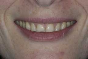
Pre-op smile (left)
Figure 2 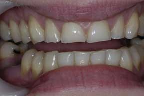
Pre-op retracted (right)
Figure 3 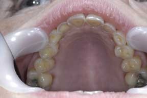
Pre-op occlusal view showing erosion (left)
Figure 4 
Occlusal view preparations (right)
Figure 5 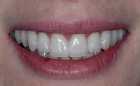
Post-op smile (left)
Figure 6 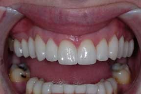
Post-op retracted (right)
Figure 7 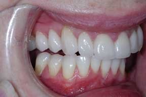
Right hand excursion (left)
Figure 8 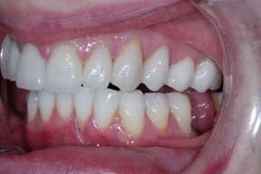
Left hand excursion (right)
-
4
Bone loss. Radiographic examination can show loss of lamina dura and both vertical and horizontal bone loss as well as caries. Angular bony defects may be particularly indicative of jiggling forces in the presence of plaque.7


For all these reasons the dynamic situation has to be assessed and will involve centric relation and centric occlusion assessment as well as excursive movements.
From balanced articulation (dentures) to group function, and canine guidance many excursive schemes exist. Cr/co discrepancies as well as the presence of posterior working and non working interferences should be evaluated as occlusal slides invariably result in destructive interferences and these can cause many of the symptoms outlined above.
The goal of occlusal therapy is not to stop parafunctional activity. It is to put the patient at a mechanical disadvantage during the parafunction thus minimising the stress to the masticatory system.
All the above, together with a full set of clinical photographs (invaluable documentation for medico legal considerations), enables a thorough diagnosis and a comprehensive treatment plan to be formulated before it is presented to the patient. All treatment plan options should be discussed with the patient to allow informed consent.
Articulated study models mounted in centric relation should be used to perform a trial equilibration to assess the amount of enamel adjustment required to achieve cr/co coincidence. The advent of modern bonding additive procedures such as canine bonding can allow such posterior disclussion if the patient does not have aesthetic concerns.
Patient discussions can involve composite mock-ups and digital imaging (see case 2). These may address the aesthetic concerns but not the functional issues. A diagnostic wax up allows not only confirmation of the proposed functional aesthetic scheme on a semi-adjustable articulator and then intra-oral refinement but also prototypes to allow the patient to visualise the changes to meet their requirements.
Conjunctive orthodontic therapy can often achieve many of the functional requirements outlined above and thus minimise the extent of tooth preparation. However, if the patient is unwilling to accept this, a restorative solution is one of five choices (see Table 1).
Even heavily filled teeth can be restored with veneers — these may be anywhere between 180 degree with incisal coverage and 360 degree coverage depending on remaining tooth tissue — all margins should however be placed on sound tooth enamel and not pre-existing restorations.8
Technique
See part one in this series — Aesthetic changes with four anterior units (BDJ 2006; 200: 135–138).
Case descriptions
For all the cases illustrated in this article, the dentistry was performed to satisfy the patient's desires for aesthetic changes to their teeth and not primarily to change their occlusal scheme. If the aesthetic changes are to be made then these changes need to be completed in a logical, repeatable and organised manner. This can best be achieved by using a recognised occlusal scheme5 and corrections to their occlusal scheme should be incorporated. Without this the likelihood of de-bonds and fractures will only increase.
In all cases all options available were discussed. These included no treatment, bleaching only, direct resin bonding or porcelain veneers to correct size shape discrepancy and orthodontics. All necessary hygiene treatment was completed for these patients and active caries or leaking restorations addressed prior to cosmetic treatment.
Case 1
This patient requested options for her wearing anterior, discoloured teeth following a history of bulimia. She had the associated characteristic palatal erosion which was through the enamel and into the dentine, as well as loss of canine guidance. The patient wanted to smile with confidence again and upon discussion of her available options she decided upon 10 upper porcelain laminate veneers with 360 degree coverage where required and bleaching of her lower teeth. As well as restoring the loss of incisal length, canine protected occlusion was achieved. All the exposed dentine was protected with virtually no preparation of the palatal aspects of the anterior teeth and only very minimal preparation of the occlusal surfaces of the premolars. No increase in the occlusal vertical dimension was required (Figs 1,2,3,4,5,6,7,8).
Case 2
Despite previous bleaching the patient wanted to improve her smile. She was unaware of her congenitally missing lateral incisors and believed further bleaching could improve the appearance of her teeth. Imaging and mock-ups allow the patient to see her aesthetic options.9 Completion of a comprehensive examination including co-diagnosis allowed the association between the abfraction, incisal notching (which she had always believed was present from a historical accident and subsequent dental adjustment) to be discussed with the patient. The patient elected to have a 10 unit upper smile lift with bleaching of her lower teeth (Figs 9,10,11, 12,13,14,15,16,17,18,19,20).

Pre-op smile (left)

Retracted pre-op smile (right)

Close up pre-op smile (left)

Mock up of canines into laterals (right)

Mock up of four upper anteriors (left)

Imaged face (right)

Prototypes (left)

Prototypes close up (right)

Occlusal view of prototypes (left)

Post-op smile (right)

Retracted post-op (left)

Final occlusal view matches prototypes (right)
Case 3
Having had a previous history of a high sugar diet, this patient presented requesting a complete smile make-over. As well as disliking the colour he disliked the multiple diastemas.10 He was unaware of the anterior cross bite and the non-working interference on tooth 16 and the associated angular bony defect.11 Via appropriate conservative preparations his cosmetic and our functional concerns could be resolved (Figs 21,22,23,24,25,26).

Pre-op smile (left)

Pre-op retracted: note cross bite (right)

Angular bony defect (right)

Preps (right)

Post-op smile (right)

Retracted post-op (right)
Discussion
Immediate value for the patient is determined by the aesthetics of the case, the comfort of the reconstruction and the care in which the dentistry was delivered. Long-term value will be determined by longevity and continuing patient satisfaction.
References
Orr C . 12 steps to smile design 1: Macroaesthetic elements. Aest Imp Dent 2005; 7.1: 16–22.
Dunne SM, Millar BJ . A longitudinal study of the clinical performance of porcelain veneers. Br Dent J 1993; 175: 317–322.
Dumfahrt H, Schaffer H . Porcelain laminate veneers. A retrospective evaluation after 1 to 10 years of service: Part II — Clinical results. Int J Prosthodont 2000; 13: 9–18.
American Academy of Cosmetic Dentistry (AACD) A guide to accreditation photography 2004. AACD.COM
Dawson PE . Evaluation, diagnosis and treatment of occlusal problems. pp280–285. St Louis, MO: CV Mosby, 1989.
Dawson P . Position paper regarding diagnosis, management, and treatment of temporomandibular disorders. The American Equilibration Society. J Prosthet Dent 1999; 81: 174–178.
Ericsson I . The combined effects of plaque and physical stress on periodontal tissues. J Clin Periodontol 1986; 13: 918–922.
Edelhoff D, Sorensen JA . Tooth structure removal associated with various preparation designs for anterior teeth. J Prosthet Dent 2002; 87: 503–509.
Millar BJ, Taylor NG . Lateral thinking: the management of missing upper lateral incisors. Br Dent J 1995; 179: 99–106.
Tarnow DP, Magner AW, Fletcher P . The effect of the distance from the contact point to the crest of bone on the presence or absence of the interproximal papilla. J Periodontol 1992; 63: 995–996.
Schuyler CH . Factors contributing to traumatic Occlusion. J Prosthetic Dent 1961; II: 708–711.
Rufenacht CR . Fundamentals of aesthetics. UK: Quintessence, 1990.
Gurel G . The science and art of porcelain laminate veneers. UK: Quintessence, 2003.
Acknowledgements
Many thanks to Luke Barnett Dental Ceramic Specialists in Watford for all the porcelain work shown. We are also indebted to Roy Higson for providing us with an occlusal learning pathway (www.ipsoseminars.org) and his continued support.
Author information
Authors and Affiliations
Corresponding author
Additional information
Refereed Paper
Rights and permissions
About this article
Cite this article
Bloom, D., Padayachy, J. Smile lifts — A functional and aesthetic perspective. Br Dent J 200, 199–203 (2006). https://doi.org/10.1038/sj.bdj.4813252
Published:
Issue Date:
DOI: https://doi.org/10.1038/sj.bdj.4813252
