
Key Points
-
Most red or hyperpigmented lesions in the mouth are inconsequential.
-
However, cancer and some systemic diseases may present in this way.
-
Most red lesions are inflammatory or atrophic but erthythroplasia is potentially malignant.
-
Most hyperpigmented lesions are racial or due to embedded material (eg amalgam tattoo) but malignant and systemic disease can present in this way.
-
Biopsy may be indicated.
Key Points
Oral medicine
-
1
Aphthous and other common ulcers
-
2
Mouth ulcers of more serious connotation
-
3
Dry mouth and disorders of salivation
-
4
Oral malodour
-
5
Oral white patches
-
6
Oral red and hyperpigmented patches
-
7
Orofacial sensation and movement
-
8
Orofacial swellings and lumps
-
9
Oral cancer
-
10
Orofacial pain
Abstract
This series provides an overview of current thinking in the more relevant areas of oral medicine for primary care practitioners, written by the authors while they were holding the Presidencies of the European Association for Oral Medicine and the British Society for Oral Medicine, respectively. A book containing additional material will be published. The series gives the detail necessary to assist the primary dental clinical team caring for patients with oral complaints that may be seen in general dental practice. Space precludes inclusion of illustrations of uncommon or rare disorders, or discussion of disorders affecting the hard tissues. Approaching the subject mainly by the symptomatic approach — as it largely relates to the presenting complaint — was considered to be a more helpful approach for GDPs rather than taking a diagnostic category approach. The clinical aspects of the relevant disorders are discussed, including a brief overview of the aetiology, detail on the clinical features and how the diagnosis is made. Guidance on management and when to refer is also provided, along with relevant websites which offer further detail.
Red and pigmented lesions
This article covers red lesions followed by hyperpigmentation.
Red oral lesions
Red oral lesions are commonplace and usually associated with inflammation in, for example, mucosal infections. However, red lesions can also be sinister by signifying severe dysplasia in erythroplasia, or malignant neoplasms (Table 1).
Geographic tongue (erythema migrans)
Geographic tongue (Fig. 1) is a very common condition and cause of sore tongue, affecting at least 1-2% of patients. There is a genetic background, and often a family history. Many patients with a fissured tongue (scrotal tongue) also have geographic tongue. Erythema migrans is associated with psoriasis in 4% and the histological appearances of both conditions are similar. Some patients have atopic allergies such as hay fever and a few relate the symptoms to various foods. A few have diabetes mellitus.

Geographic tongue
Clinical features
Geographic tongue typically involves the dorsum of the tongue, sometimes the ventrum. It is often asymptomatic but a small minority of patients complain of soreness; these patients are virtually invariably middle-aged. If sore, this may be noted especially with acidic foods (for example tomatoes or citrus fruits) or cheese.
There are irregular, pink or red depapillated maplike areas, which change in shape, increase in size, and spread or move to other areas sometimes within hours (Figs 2 and 3).

Geographic tongue

Geographic tongue

Candida-associated denture stomatitis

Median rhomboid glossitis

Erythematous candidosis
The red areas are often surrounded by distinct yellowish slightly raised margins. There is increased thickness of the intervening filiform papillae.
Diagnosis

The diagnosis of geographic tongue is clinical mainly from the history of a migrating pattern and the characteristic clinical appearance. Blood examination may rarely be necessary to exclude diabetes, or anaemia if there is confusion with a depapillated tongue of glossitis.
Management
Reassurance remains the best that can be given. Zinc sulphate 200mg three times daily for three months or a topical rinse with 7% salicylic acid in 70% alcohol are advocated by some and may occasionally help.
Patient information and websites
http://www.usc.edu/hsc/dental/opath/Cards/GeographicTongue.html
Denture-related stomatitis (Denture-induced stomatitis; denture sore mouth; chronic erythematous candidosis)
Denture-related stomatitis consists of mild inflammation of the mucosa beneath a denture — usually a complete upper denture. This is a common condition, mainly of the middle-aged or elderly, more prevalent in women than men.
Aetiopathogenesis
Dental appliances (mainly dentures) especially when worn throughout the night, or a dry mouth, favour development of this infection. It is not caused by allergy to the dental material (if it were, it would affect mucosae other than just that beneath the appliance).
However, it is still not clear why only some denture wearers develop denture-related stomatitis, since most patients appear otherwise healthy.
Dentures can produce a number of ecological changes; the oral flora may be altered and plaque collects between the mucosal surface of the denture and the palate.
The accumulation of microbial plaque (bacteria and/or yeasts) on and attached to the fitting surface of the denture and the underlying mucosa produces an inflammatory reaction. When candida is involved, the more common terms 'candida-associated denture stomatitis', 'denture-induced candidosis' or 'chronic erythematous candidosis' are used.
In addition, the saliva that is present between the maxillary denture and the mucosa may have a lower pH than usual. Denture-related stomatitis is sometimes associated also with various bacteria but is not exclusively associated with infection, and occasionally mechanical irritation is at play.
Clinical features
The characteristic presenting features of denture-related stomatitis are chronic erythema and oedema of the mucosa that contacts the fitting surface of the denture (Fig. 2). Uncommon complications include:
-
Angular stomatitis
-
Papillary hyperplasia in the vault of the palate.
Classification
Denture-related stomatitis has been classified into three clinical types (Newton's types), increasing in severity:
-
A localised simple inflammation or a pinpoint hyperaemia (Type I)
-
An erythematous or generalised simple type presenting as more diffuse erythema involving part of or the entire, denture-covered mucosa (Type II)
-
A granular type (inflammatory papillary hyperplasia) commonly involving the central part of the hard palate and the alveolar ridge (Type III).
Diagnosis
Denture-related stomatitis is a clinical diagnosis although it may be confirmed by microbiological investigations. In addition haematological and biochemical investigations may be appropriate to identify any underlying predisposing factors such as nutritional deficiencies, anaemia and diabetes mellitus in patients unresponsive to conventional management.
Management

The denture plaque and fitting surface is infested with micro-organisms, most commonly Candida albicans and therefore, to prevent recurrence, dentures should be left out of the mouth at night, and stored in an appropriate antiseptic which has activity against yeasts (Table 2).
Cleansers containing alkaline hypochlorites, disinfectants, or yeast lytic enzymes are most effective against candida. Denture soak solution containing benzoic acid is taken up into the acrylic resin and can completely eradicate C.albicans from the denture surface. Chlorhexidine gluconate can also eliminate C.albicans on the denture surface and a mouthwash can reduce the palatal inflammation.
The mucosal infection is eradicated by brushing the palate with chlorhexidine mouthwash or gel, and using miconazole gel, nystatin pastilles, amphotericin lozenges or fluconazole, administered concurrently with an oral antiseptic such as chlorhexidine which has antifungal activity.
Patient information and website
http://www.emedicine.com/derm/topic642.htm
Neoplastic lesions; red neoplasms include:
-
Peripheral giant cell tumours
-
Angiosarcomas such as Kaposi's sarcoma—a common neoplasm in HIV/AIDS, appears in the mouth as red or purplish areas or nodules especially seen in the palate
-
Squamous cell carcinomas
-
Wegener's granulomatosis.
Vascular anomalies (angiomas and telangiectasia) include:
-
Dilated lingual veins (varices) may be conspicuous in normal elderly persons
-
Haemangiomas are usually small isolated developmental anomalies, or hamartomas (Figs 7,8,9)
-
Telangiectasias — dilated capillaries — may be seen after irradiation and in disorders such as hereditary haemorrhagic telangiectasia and systemic sclerosis (Fig. 10)
-
Angiomas are benign and usually congenital (Figs 7,8,9,10). In general most do not require any active treatment unless symptoms develop, in which case they can be treated by injection of sclerosing agents, cryosurgery, laser excision or surgical excision.
Figure 7 
Vascular hamartoma (haemangioma) tongue
Figure 8 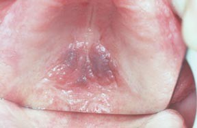
Vascular hamartoma (haemangioma, palate)
Figure 9 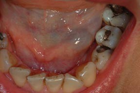
Haemangioma in floor of mouth
Figure 10 
Telangiectasia, lips and tongue


Vesiculobullous disorders

Erythema multiforme, pemphigoid and pemphigus may present as red lesions (see article two), especially localised oral purpura, which presents with blood blisters (Fig. 11). Specialist referral is usually indicated.

Angina bullosa haemorrhagica
Reactive lesions
Reactive lesions that can be red are usually persistent soft lumps (Figs 12 and 13) which include:
-
Pyogenic granulomas
-
Peripheral giant cell granulomas

Pyogenic granuloma, lower lip

Pyogenic epulis
Specialist referral is usually indicated.
Atrophic lesions
The most important red lesion is erythroplasia, since it is often dysplastic (see below). Geographic tongue also causes red lesions (see above), desquamative gingivitis is a frequent cause of red gingivae, almost invariably caused by lichen planus or pemphigoid, and iron or vitamin deficiency states may cause glossitis (Fig. 14) or other red lesions.

Atrophic glossitis
Erythroplakia (erythroplasia)
Erythroplasia is a rare condition defined as 'any lesion of the oral mucosa that presents as bright red velvety plaques which cannot be characterised clinically or pathologically as any other recognisable condition'.
Mainly seen in elderly males, it is far less common than leukoplakia, but far more likely to be dysplastic or undergo malignant transformation.
Clinical features
Erythroplakia is seen most commonly on the soft palate, floor or mouth or buccal mucosa. Some erythroplakias are associated with white patches, and are then termed speckled leukoplakia (Fig. 15).

Erythroplasia in soft palate complex
Diagnosis
Biopsy to assess the degree of epithelial dysplasia and exclude a diagnosis of carcinoma.
Prognosis
Erythroplasia has areas of dysplasia, carcinoma in situ, or invasive carcinoma in most cases. Carcinomas are seen 17 times more often in erythroplakia than in leukoplakia and these are therefore the most potentially malignant of all oral mucosal lesions.
Management
Erythroplastic lesions are usually (at least 85%) severely dysplastic or frankly malignant. Any causal factor such as tobacco use should be stopped, and lesions removed. There is no hard evidence as to the ideal frequency of follow-up, but it has been suggested that patients with mucosal potentially malignant lesions be re-examined within one month, at three months, at six months, at 12 months and annually thereafter.
Purpura
This presents as bleeding into the skin and mucosa and is usually caused by trauma. Occasional small petechiae are seen at the occlusal line in perfectly healthy people.
Thrombocytopenia can result in red or brown pinpoint lesions (petechiae) or diffuse bruising (ecchymoses) at sites of trauma, such as the palate. Suction (eg fellatio) may produce bruising in the soft palate). Localised oral purpura or angina bullosa haemorrhagica is an idiopathic, fairly common cause of blood blisters, often in the soft palate, in older persons (Fig. 11). Sometimes the use of a corticosteroid inhaler precipitates this.
Diagnosis of red lesions
Diagnosis of red lesions is mainly clinical but lesions should also be sought elsewhere, especially on the skin or other mucosae.
It may be necessary to take a blood picture (including blood and platelet count), and assess haemostatic function or exclude haematinic deficiencies. Other investigations needed may include other haematological tests and/or biopsy or imaging.
Management
Treatment is usually of the underlying cause, or surgery.
Hyperpigmentation
Oral mucosal discolouration may be superficial (extrinsic) or due to deep (intrinsic — in or beneath mucosa) causes and ranges from brown to black.


Extrinsic discolouration is rarely of consequence and is usually caused by:
-
Habits such as tobacco or betel use
-
Coloured foods or drinks, (such as liquorice, beetroot, red wine, coffee, tea)
-
Drugs (such as chlorhexidine, iron salts, crack cocaine, minocycline, bismuth subsalicylate, and lansoprazole).
Black hairy tongue
This is one extrinsic type of discolouration seen especially in patients on a soft diet, smokers, and those with dry mouth or poor oral hygiene (Fig. 16).

Black hairy tongue
The best that can usually be done is to avoid the cause where known, and to advise the patient to brush the tongue or use a tongue-scraper.
Intrinsic discolouration
This may have much more significance (Table 3). Localised areas of pigmentation may be caused mainly by:
-
Amalgam tattoo (embedded amalgam). Typically this is a single blue-black macule in the mandibular gingiva close to the scar of an apicectomy (Figs 17 and 18) or where amalgam has accidentally been introduced into a wound, is painless, and does not change in size or colour. A lesion suspected to be an amalgam tattoo is best radiographed first to see if there is radio-opaque material present, though not all are radio-opaque. If the lesion is not radio-opaque, it is best biopsied to exclude naevi or melanoma. Similar lesions can be caused by other foreign bodies (eg graphite tattoo), local irritation or inflammation.
Figure 17 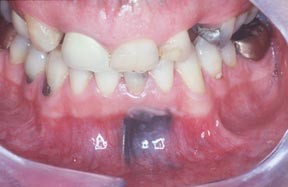
Amalgam tattoo
Figure 18 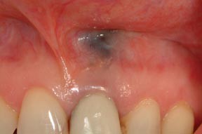
Amalgam tattoo
-
Naevi are blue-black often papular lesions formed from increased melanin-containing cells (naevus cells) seen particularly on the palate. They are best removed to exclude melanoma.
-
Pigmentary incontinence may be seen in some inflammatory lesions such as lichen planus, especially in smokers (Fig. 20).
Figure 20 
Smoking-induced melanosis, buccal mucosa
-
Melanotic macules are usually flat single brown, collections of melanin-containing cells, seen particularly on the vermilion border of the lip and on the palate (Fig. 19). They are best removed to exclude melanoma.
Figure 19 
Melanotic macule, lower labial mucosa
-
Malignant melanoma is rare, seen usually in the palate or maxillary gingivae. Features suggestive of malignancy include a rapid increase in size, change in colour, ulceration, pain, the occurrence of satellite pigmented spots or regional lymph node enlargement. Incisional biopsy to confirm the diagnosis followed by radical excision is indicated.
-
Kaposi's sarcoma is usually a purple lesion seen mainly in the palate or gingival of HIV-infected and other immunocompromised persons.


, , ,
Generalised pigmentation, often mainly affecting the gingivae, is common in persons of colour, and is racial and due to melanin. Seen mainly in black and ethnic minority groups it can also be noted in some fairly light-skinned people (Fig. 21). Such pigmentation may be first noted by the patient in adult life and then incorrectly assumed to be acquired.

Racial pigmentation
In all other patients with widespread intrinsic pigmentation, systemic causes should be excluded. These may include:
-
Tobacco, which can also cause intrinsic hyperpigmentation (smoker's melanosis)
-
Antimalarials, oral contraceptive pill, anticonvulsants, minocycline, phenothiazines, gold, busulphan and other drugs
-
Heavy metals (such as mercury, lead and bismuth) not used therapeutically now, rarely cause industrial exposure etc
-
Pregnancy
-
Hypoadrenalism (Addison's disease). Hyperpigmentation in this is generalised but most obvious in normally pigmented areas (eg the nipples, genitalia), skin flexures, and sites of trauma. The mouth may show patchy hyperpigmentation. Patients also typically have weakness, weight loss, and hypotension.
Diagnosis
The nature of oral hyperpigmentation can sometimes only be established after further investigation.

In patients with localised hyperpigmentation, in order to exclude melanoma, radiographs may be helpful (they can sometimes show a foreign body) and biopsy may be indicated, particularly where there is a solitary raised lesion, a rapid increase in size, change in colour, ulceration, pain, evidence of satellite pigmented spots or regional lymph node enlargement. If early detection of oral melanomas is to be achieved, all pigmented oral cavity lesions should be viewed with suspicion. The consensus of opinion is that a lesion with clinical features as above seriously suggestive of malignant melanoma, are best biopsied at the time of definitive operation.
In patients with generalised or multiple hyperpigmentation, specialist referral is indicated.
Management
Management is of the underlying condition.
Author information
Authors and Affiliations
Corresponding author
Additional information
Refereed Paper
Rights and permissions
About this article
Cite this article
Scully, C., Felix, D. Oral Medicine — Update for the dental practitioner Red and pigmented lesions. Br Dent J 199, 639–645 (2005). https://doi.org/10.1038/sj.bdj.4813017
Published:
Issue Date:
DOI: https://doi.org/10.1038/sj.bdj.4813017
This article is cited by
-
Palatal Erythema with Histological Psoriasiform Pattern: An Enigmatic Oral Finding Shared by a Range of Conditions
Head and Neck Pathology (2020)
-
Toluidine blue color perception in identification of oral mucosal lesions
Clinical Oral Investigations (2011)
