Thank you for visiting nature.com. You are using a browser version with limited support for CSS. To obtain
the best experience, we recommend you use a more up to date browser (or turn off compatibility mode in
Internet Explorer). In the meantime, to ensure continued support, we are displaying the site without styles
and JavaScript.
In this Tools of the Trade article, Isomursu (Ivaska lab) describes a new method for dynamic micropatterning, which enables investigation of cell adhesion and migration on substrates that mimic different extracellular matrix environments.
In this Tools of the Trade article, Venkova and Popard (Piel lab) discuss recent updates to the fluorescence exclusion method that now enable simultaneous measurement of cellular and nuclear size as well as investigation of small prokaryotic cells.
Reversible S-palmitoylation regulates gasdermin D cleavage, membrane translocation and pore formation to control pyroptosis following bacterial infection.
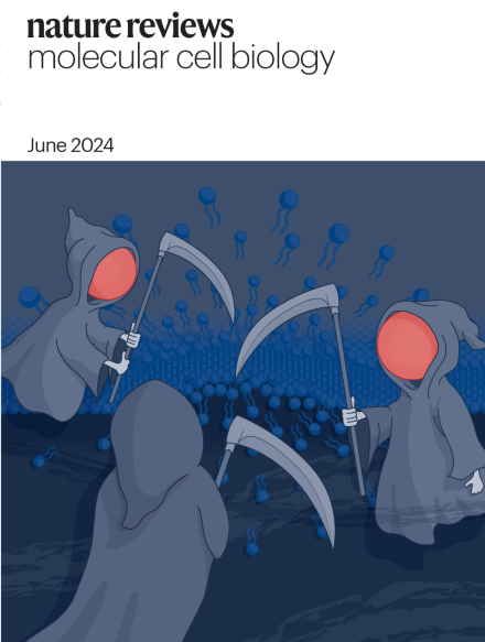
Ferroptosis is a non-apoptotic, iron-dependent cell death mechanism driven by plasma membrane lipid peroxidation and subsequent plasma membrane rupture. Various cellular compartments and organelles contribute to regulating susceptibility to ferroptosis. This regulation involves a plethora of mechanisms centred on iron metabolism and storage, lipid metabolism, and redox balance.
The prevailing challenge in live-cell fluorescence microscopy is capturing intra-cellular dynamics while preserving cell viability. Alongside developments of microscopy hardware, computational methods — especially those based on machine learning — are powerful tools to improve the signal-to-noise ratio, spatial resolution, temporal resolution and multi-colour capacity of live-cell imaging.
L. Villiger, J. Joung et al. review CRISPR applications for programmable editing of the genome, epigenome and transcriptome. They discuss how CRISPR–Cas systems can be optimized to further improve editing specificity and efficiency and highlight a multitude of applications in basic biological research and for changing clinical practice.
Protein S-acylation is involved in many pathophysiological processes. Here, Mesquita et al. discuss the structure, function and regulation of S-acylation and deacylation enzymes and describe how this post-transcriptional modification precisely controls protein–cell membrane interactions. Potential therapeutic applications of S-acylation are also highlighted.