


« Prev Next »

Replication and Distribution of DNA during Meiosis
Like mitosis, meiosis is a form of eukaryotic cell division. However, these two processes distribute genetic material among the resulting daughter cells in very different ways. Mitosis creates two identical daughter cells that each contain the same number of chromosomes as their parent cell. In contrast, meiosis gives rise to four unique daughter cells, each of which has half the number of chromosomes as the parent cell. Because meiosis creates cells that are destined to become gametes (or reproductive cells), this reduction in chromosome number is critical — without it, the union of two gametes during fertilization would result in offspring with twice the normal number of chromosomes!
Apart from this reduction in chromosome number, meiosis differs from mitosis in yet another way. Specifically, meiosis creates new combinations of genetic material in each of the four daughter cells. These new combinations result from the exchange of DNA between paired chromosomes. Such exchange means that the gametes produced through meiosis exhibit an amazing range of genetic variation.
Finally, unlike mitosis, meiosis involves two rounds of nuclear division, not just one. Despite this fact, many of the other events of meiosis are similar to those that occur in mitosis. For example, prior to undergoing meiosis, a cell goes through an interphase period in which it grows, replicates its chromosomes, and checks all of its systems to ensure that it is ready to divide. Like mitosis, meiosis also has distinct stages called prophase, metaphase, anaphase, and telophase. A key difference, however, is that during meiosis, each of these phases occurs twice — once during the first round of division, called meiosis I, and again during the second round of division, called meiosis II.
What happens during meiosis I?
As previously mentioned, the first round of nuclear division that occurs during the formation of gametes is called meiosis I. It is also known as the reduction division because it results in cells that have half the number of chromosomes as the parent cell. Meiosis I consists of four phases: prophase I, metaphase I, anaphase I, and telophase I.
Prophase I
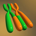
Figure 1: Recombination is the exchange of genetic material between homologous chromosomes.
During prophase I, the chromosomes condense and become visible inside the nucleus. Because each chromosome was duplicated during the S phase that occurred just before prophase I, each now consists of two sister chromatids joined at the centromere. This arrangement means that each chromosome has the shape of an X.
Once this chromosomal condensation has occurred, the members of each chromosome pair (called homologous chromosomes, because they are similar in size and contain similar genes), align next to each other. At this point, the two chromosomes in each pair become tightly associated with each other along their lengths in a process called synapsis. Then, while the homologous chromosomes are tightly paired, the members of each pair trade adjacent bits of DNA in a process called crossing over, also known as recombination (Figure 1). This trading of genetic material creates unique chromosomes that contain new combinations of alleles.
At the end of prophase I, the nuclear membrane finally
begins to break down. Outside the nucleus, the spindle grows out from
centrosomes on each side of the cell. As in mitosis, the microtubules of the
spindle are responsible for moving and arranging the chromosomes during
division.
Metaphase I
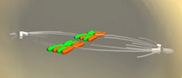
Figure 2: Near the end of metaphase I, the homologous chromosomes align on the metaphase plate.
At the start of metaphase I, microtubules emerge from the spindle and attach to the kinetochore near the centromere of each chromosome. In particular, microtubules from one side of the spindle attach to one of the chromosomes in each homologous pair, while microtubules from the other side of the spindle attach to the other member of each pair. With the aid of these microtubules, the chromosome pairs then line up along the equator of the cell, termed the metaphase plate (Figure 2).
Anaphase I
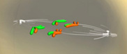
Figure 3: During anaphase I, the homologous chromosomes are pulled toward opposite poles of the cell.
During anaphase I, the
microtubules disassemble and contract; this, in turn, separates the homologous
chromosomes such that the two chromosomes in each pair are pulled toward opposite
ends of the cell (Figure 3). This separation means that each of the daughter
cells that results from meiosis I will have half the number of chromosomes of
the original parent cell after interphase. Also, the sister chromatids in each chromosome
still remain connected. As a result, each chromosome maintains its X-shaped
structure.
Telophase I
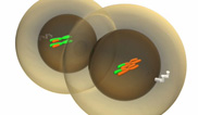
Figure 4: Telophase I results in the production of two nonidentical daughter cells, each of which has half the number of chromosomes of the original parent cell.
As the new chromosomes reach the spindle during telophase I, the cytoplasm organizes itself and divides in two. There are now two cells, and each cell contains half the number of chromosomes as the parent cell. In addition, the two daughter cells are not genetically identical to each other because of the recombination that occurred during prophase I (Figure 4).
Interkinesis
At this point, the first division of meiosis is complete. The cell now rests
for a bit before beginning the second meiotic division. During this period,
called interkinesis, the
nuclear membrane in each of the two cells reforms around the chromosomes. In
some cells, the spindle also disintegrates and the chromosomes relax (although
most often, the spindle remains intact).
It is important to note, however, that no chromosomal duplication occurs during this stage.
What happens during meiosis II?
During meiosis II, the two cells once again cycle through four phases of division. Meiosis II is sometimes referred to as an equational division because it does not reduce chromosome number in the daughter cells — rather, the daughter cells that result from meiosis II have the same number of chromosomes as the "parent" cells that enter meiosis II. (Remember, these "parent" cells already have half the number of chromosomes of the original parent cell thanks to meiosis I.)
Prophase II
As prophase
II begins, the chromosomes once again condense into tight structures, and
the nuclear membrane disintegrates. In addition, if the spindle was disassembled
during interkinesis, it reforms at this point in time.
Metaphase II
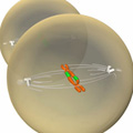
Figure 5: During metaphase II, the chromosomes align along the cell's equatorial plate.
The events of metaphase II are similar to those of mitotic metaphase — in both
processes, the chromosomes line up along the cell's equatorial plate, also
called the metaphase plate, in preparation for their eventual separation
(Figure 5).
Anaphase II
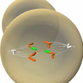
Figure 6: Anaphase II involves separation of the sister chromatids.
During anaphase
II, microtubules from each spindle attach to each sister chromatid at the
kinetochore. The sister chromatids then separate, and the microtubules pull
them to opposite poles of the cell. As in mitosis, each chromatid is now
considered a separate chromosome (Figure 6). This means that the cells that
result from meiosis II will have the same number of chromosomes as the "parent"
cells that entered meiosis II.
Telophase II
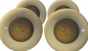
Figure 7: Telophase II results in the production of four daughter cells.
Finally, in telophase II, nuclear
membranes reform around the newly separated chromosomes, which relax and fade
from view. As soon as the cytoplasm divides, meiosis is complete. There are now
four daughter cells — two from each of the two cells that entered meiosis II —
and each daughter cell has half the normal number of chromosomes (Figure 7).
Each also contains new mixtures of genes within its chromosomes, thanks to
recombination during meiosis I.
Why is meiosis important?
Meiosis is important because it ensures that all
organisms produced via sexual reproduction contain the correct number of
chromosomes. Meiosis also produces genetic variation by way of the process of
recombination. Later, this variation is increased even further when two gametes
unite during fertilization, thereby creating offspring with unique combinations
of DNA. This constant mixing of parental DNA in sexual reproduction helps fuel
the incredible diversity of life on Earth.
Further Exploration
eBooks
This page appears in the following eBook


