


« Prev Next »

Mitosis
Mitosis is the process in which a eukaryotic cell nucleus splits in two, followed by division of the parent cell into two daughter cells. The word "mitosis" means "threads," and it refers to the threadlike appearance of chromosomes as the cell prepares to divide. Early microscopists were the first to observe these structures, and they also noted the appearance of a specialized network of microtubules during mitosis. These tubules, collectively known as the spindle, extend from structures called centrosomes — with one centrosome located at each of the opposite ends, or poles, of a cell. As mitosis progresses, the microtubules attach to the chromosomes, which have already duplicated their DNA and aligned across the center of the cell. The spindle tubules then shorten and move toward the poles of the cell. As they move, they pull the one copy of each chromosome with them to opposite poles of the cell. This process ensures that each daughter cell will contain one exact copy of the parent cell DNA.
What Are the Phases of Mitosis?
Mitosis consists of five morphologically distinct phases: prophase, prometaphase, metaphase, anaphase, and telophase. Each phase involves characteristic steps in the process of chromosome alignment and separation. Once mitosis is complete, the entire cell divides in two by way of the process called cytokinesis (Figure 1).
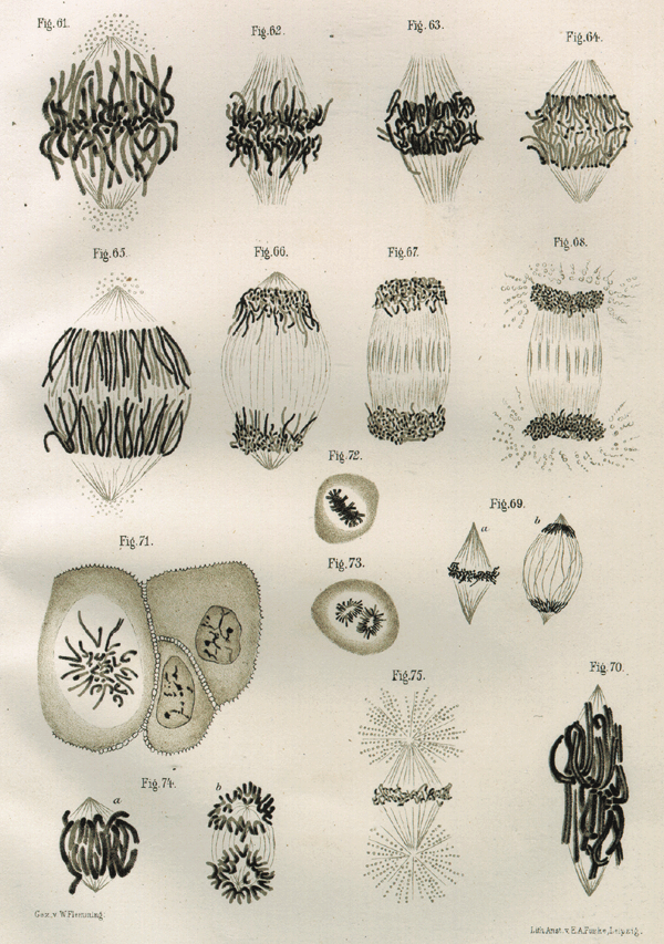
Figure 1: Drawing of chromosomes during mitosis by Walther Flemming, circa 1880
This illustration is one of more than one hundred drawings from Flemming's \"Cell Substance, Nucleus, and Cell Division.\" Flemming repeatedly observed the different forms of chromosomes leading up to and during cytokinesis, the ultimate division of one cell into two during the last stage of mitosis.
© 2001 Nature Publishing Group Paweletz, N. Walther Flemming: pioneer of mitosis research. Nature Reviews Molecular Cell Biology 2, 72 (2001). All rights reserved. 
What Happens during Prophase?
Prophase is the first stage in mitosis, occurring after the conclusion of the G2 portion of interphase. During prophase, the parent cell chromosomes — which were duplicated during S phase — condense and become thousands of times more compact than they were during interphase. Because each duplicated chromosome consists of two identical sister chromatids joined at a point called the centromere, these structures now appear as X-shaped bodies when viewed under a microscope. Several DNA binding proteins catalyze the condensation process, including cohesin and condensin. Cohesin forms rings that hold the sister chromatids together, whereas condensin forms rings that coil the chromosomes into highly compact forms.
The mitotic spindle also begins to develop during prophase. As the cell's two centrosomes move toward opposite poles, microtubules gradually assemble between them, forming the network that will later pull the duplicated chromosomes apart.
What Happens during Prometaphase?
When prophase is complete, the cell enters prometaphase — the second stage of mitosis. During prometaphase, phosphorylation of nuclear lamins by M-CDK causes the nuclear membrane to break down into numerous small vesicles. As a result, the spindle microtubules now have direct access to the genetic material of the cell.
Each microtubule is highly dynamic, growing outward from the centrosome and collapsing backward as it tries to locate a chromosome. Eventually, the microtubules find their targets and connect to each chromosome at its kinetochore, a complex of proteins positioned at the centromere. The actual number of microtubules that attach to a kinetochore varies between species, but at least one microtubule from each pole attaches to the kinetochore of each chromosome. A tug-of-war then ensues as the chromosomes move back and forth toward the two poles.
What Happens during Metaphase and Anaphase?
As prometaphase ends and metaphase begins, the chromosomes align along the cell equator. Every chromosome has at least two microtubules extending from its kinetochore — with at least one microtubule connected to each pole. At this point, the tension within the cell becomes balanced, and the chromosomes no longer move back and forth. In addition, the spindle is now complete, and three groups of spindle microtubules are apparent. Kinetochore microtubules attach the chromosomes to the spindle pole; interpolar microtubules extend from the spindle pole across the equator, almost to the opposite spindle pole; and astral microtubules extend from the spindle pole to the cell membrane.
Metaphase leads to anaphase, during which each chromosome's sister chromatids separate and move to opposite poles of the cell. Enzymatic breakdown of cohesin — which linked the sister chromatids together during prophase — causes this separation to occur. Upon separation, every chromatid becomes an independent chromosome. Meanwhile, changes in microtubule length provide the mechanism for chromosome movement. More specifically, in the first part of anaphase — sometimes called anaphase A — the kinetochore microtubules shorten and draw the chromosomes toward the spindle poles. Then, in the second part of anaphase — sometimes called anaphase B — the astral microtubules that are anchored to the cell membrane pull the poles further apart and the interpolar microtubules slide past each other, exerting additional pull on the chromosomes (Figure 2).
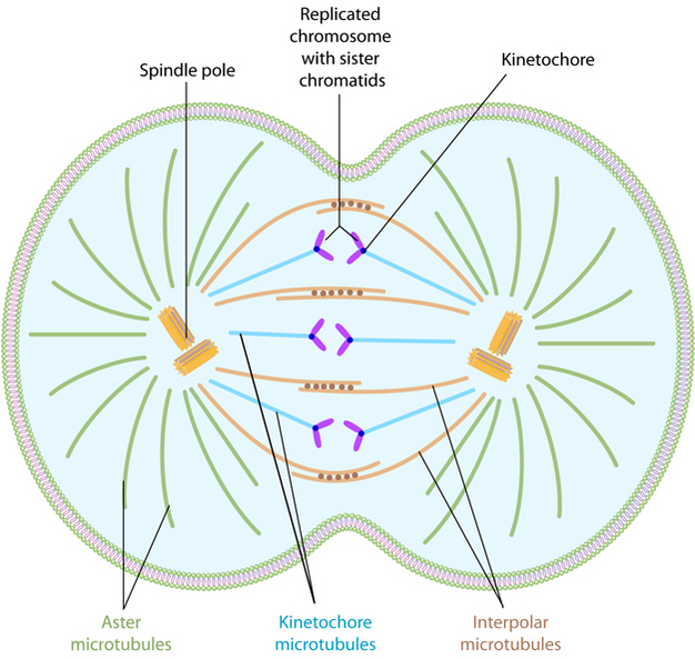
Figure 2: Types of microtubules involved in mitosis
During mitosis, several types of microtubules are active. The motor proteins associated with the interpolar microtubules drive the assembly of the spindle. Note the other types of microtubules involved in anchoring the spindle pole and pulling apart the sister chromatids.
© 2013 Nature Education All rights reserved.
What Happens during Telophase?
During telophase, the
chromosomes arrive at the cell poles, the mitotic spindle
disassembles, and
the vesicles that contain fragments of the original nuclear membrane
assemble
around the two sets of chromosomes. Phosphatases then dephosphorylate
the
lamins at each end of the cell. This dephosphorylation results in the
formation
of a new nuclear membrane around each group of chromosomes.
When Do Cells Actually Divide?
Cytokinesis is the physical process that finally splits the parent cell into two identical daughter cells. During cytokinesis, the cell membrane pinches in at the cell equator, forming a cleft called the cleavage furrow. The position of the furrow depends on the position of the astral and interpolar microtubules during anaphase.
The cleavage furrow forms because of the action of a contractile ring of overlapping actin and myosin filaments. As the actin and myosin filaments move past each other, the contractile ring becomes smaller, akin to pulling a drawstring at the top of a purse. When the ring reaches its smallest point, the cleavage furrow completely bisects the cell at its center, resulting in two separate daughter cells of equal size (Figure 3).
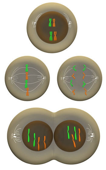
Figure 3: Mitosis: Overview of major phases
The major stages of mitosis are prophase (top row), metaphase and anaphase (middle row), and telophase (bottom row).
© 2009 Nature Education All rights reserved.
Conclusion
Mitosis
is the process of nuclear division, which occurs just prior to cell
division, or
cytokinesis. During this multistep process, cell chromosomes
condense and the spindle assembles. The duplicated chromosomes then attach to the
spindle,
align at the cell equator, and move apart as the spindle microtubules
retreat
toward opposite poles of the cell. Each set of chromosomes is then
surrounded
by a nuclear membrane, and the parent cell splits into two complete
daughter
cells.
eBooks
This page appears in the following eBook


