


« Prev Next »

Dengue Viruses
Introduction
Viruses are tiny agents that can infect a variety of living organisms, including bacteria, plants, and animals. Like other viruses, the dengue virus is a microscopic structure that can only replicate inside a host organism. Who discovered the dengue virus? How many types of dengue viruses are there, and what do we know about them? How does the dengue virus infect a cell and replicate itself? In this section, we will explore the answers to these questions.
Discovery of the Dengue Viruses
The dengue viruses are members of the genus Flavivirus in the family Flaviviridae. Along with the dengue virus, this genus also includes a number of other viruses transmitted by mosquitoes and ticks that are responsible for human diseases. Flavivirus includes the yellow fever, West Nile, Japanese encephalitis, and tick-borne encephalitis viruses.
In 1943, Ren Kimura and Susumu Hotta first isolated the dengue virus. These two scientists were studying blood samples of patients taken during the 1943 dengue epidemic in Nagasaki, Japan. A year later, Albert B. Sabin and Walter Schlesinger independently isolated the dengue virus. Both pairs of scientists had isolated the virus now referred to as dengue virus 1 (DEN-1). Is DEN-1 the only type of dengue virus?
The Dengue Serotypes
Dengue infections are caused by four closely related viruses named DEN-1, DEN-2, DEN-3, and DEN-4. These four viruses are called serotypes because each has different interactions with the antibodies in human blood serum. The four dengue viruses are similar — they share approximately 65% of their genomes — but even within a single serotype, there is some genetic variation. Despite these variations, infection with each of the dengue serotypes results in the same disease and range of clinical symptoms.
Are these four viruses all found in the same regions of the world? In the 1970s, both DEN-1 and DEN-2 were found in Central America and Africa, and all four serotypes were present in Southeast Asia. By 2004, however, the geographical distribution of the four serotypes had spread widely. Now all four dengue serotypes circulate together in tropical and subtropical regions around the world (Figure 1). The four dengue serotypes share the same geographic and ecological niche. Where did the dengue viruses first come from? Scientists hypothesize that the dengue viruses evolved in nonhuman primates and jumped from these primates to humans in Africa or Southeast Asia between 500 and 1,000 years ago.
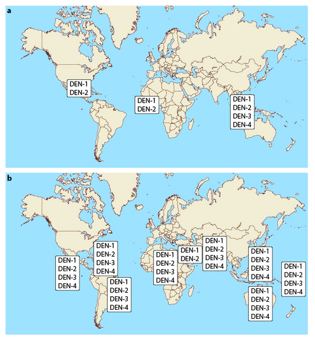
The change in distribution of dengue serotypes
The distribution of dengue serotypes in 1970 (a) and 2004 (b).
© 2014 Nature Education All rights reserved. 
After recovering from an infection with one dengue serotype, a person has immunity against that particular serotype. Does infection with one serotype protect against future dengue infections with the other serotypes? Individuals are protected from infections with the remaining three serotypes for two to three months after the first dengue infection. Unfortunately, it is not long-term protection. After that short period, a person can be infected with any of the remaining three dengue serotypes. Researchers have noticed that subsequent infections can put individuals at a greater risk for severe dengue illnesses than those who have not been previously infected.
Dengue Virus Genome and Structure
The dengue virus genome is a single strand of RNA. It is referred to as positive-sense RNA because it can be directly translated into proteins. The viral genome encodes ten genes (Figure 2). The genome is translated as a single, long polypeptide and then cut into ten proteins.
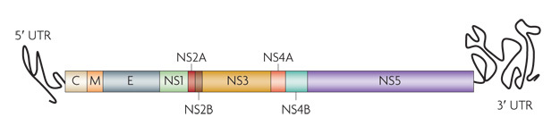
Figure 2: Dengue virus genome
The dengue virus genome encodes three structural (capsid [C], membrane [M], and envelope [E]) and seven nonstructural (NS1, NS2A, NS2B, NS3, NS4A, NS4B, and NS5) proteins.
© 2010 Nature Publishing Group Guzman, M. G. et al. Dengue: A continuing global threat. Nature Reviews Microbiology 8, S7–S16 (2010). doi:10.1038/nrmicro2460 All rights reserved.
What are the roles of these ten proteins? Three are structural proteins: the capsid (C), envelope (E), and membrane (M) proteins. Seven are nonstructural proteins: NS1, NS2A, NS2B, NS3, NS4A, NS4B, and NS5. These nonstructural proteins play roles in viral replication and assembly.
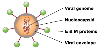
Figure 3: Dengue virus structure
The dengue virus has a roughly spherical shape. Inside the virus is the nucleocapsid, which is made of the viral genome and C proteins. The nucleocapsid is surrounded by a membrane called the viral envelope, a lipid bilayer that is taken from the host. Embedded in the viral envelope are E and M proteins that span through the lipid bilayer. These proteins form a protective outer layer that controls the entry of the virus into human cells.
© 2011 Nature Education All rights reserved.
Dengue Virus Replication and Infectious Cycle
How does the virus behave once it enters the human body? The dengue viral replication process begins when the virus attaches to a human skin cell (Figure 4). After this attachment, the skin cell's membrane folds around the virus and forms a pouch that seals around the virus particle. This pouch is called an endosome. A cell normally uses endosomes to take in large molecules and particles from outside the cell for nourishment. By hijacking this normal cell process, the dengue virus is able to enter a host cell.
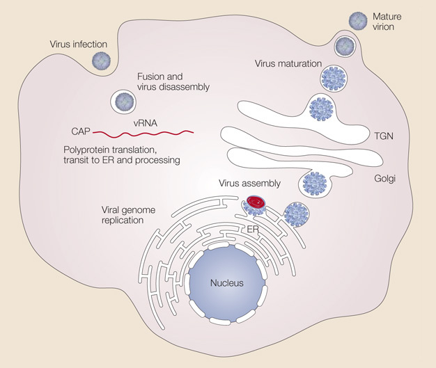
Figure 4: Dengue virus replication
The dengue virus attaches to the surface of a host cell and enters the cell by a process called endocytosis. Once deep inside the cell, the virus fuses with the endosomal membrane and is released into the cytoplasm. The virus particle comes apart, releasing the viral genome. The viral RNA (vRNA) is translated into a single polypeptide that is cut into ten proteins, and the viral genome is replicated. Virus assembly occurs on the surface of the endoplasmic reticulum (ER) when the structural proteins and newly synthesized RNA bud out from the ER. The immature viral particles are transported through the trans-Golgi network (TGN), where they mature and convert to their infectious form. The mature viruses are then released from the cell and can go on to infect other cells.
© 2005 Nature Publishing Group Mukhopadhyay, S., Kuhn, R. J., & Rossmann M. G. A structural perspective of the flavivirus life cycle. Nature Reviews Microbiology 3, 13–22 (2005). doi:10.1038/nrmicro1067 All rights reserved.
Once the virus has entered a host cell, the virus penetrates deeper into the cell while still inside the endosome. How does the virus exit the endosome, and why? Researchers have learned that two conditions are needed for the dengue virus to exit the endosome:
- The endosome must be deep inside the cell where the environment is acidic.
- The endosomal membrane must gain a negative charge.
These two conditions allow the virus envelope to fuse with the endosomal membrane, and that process releases the dengue nucleocapsid into the cytoplasm of the cell.
Once it is released into the cell cytoplasm, how does the virus replicate itself? In the cytoplasm, the nucleocapsid opens to uncoat the viral genome. This process releases the viral RNA into the cytoplasm. The viral RNA then hijacks the host cell's machinery to replicate itself. The virus uses ribosomes on the host's rough endoplasmic reticulum (ER) to translate the viral RNA and produce the viral polypeptide. This polypeptide is then cut to form the ten dengue proteins.
The newly synthesized viral RNA is enclosed in the C proteins, forming a nucleocapid. The nucleocapsid enters the rough ER and is enveloped in the ER membrane and surrounded by the M and E proteins. This step adds the viral envelope and protective outer layer. The immature viruses travel through the Golgi apparatus complex, where the viruses mature and convert into their infectious form. The mature dengue viruses are then released from the cell and can go on to infect other cells.
Summary
The dengue virus is a tiny structure that can only replicate inside a host organism. The four closely related dengue viruses — DEN-1, DEN-2, DEN-3, and DEN-4 — are found in the same regions of the world. The dengue virus is a roughly spherical structure composed of the viral genome and capsid proteins surrounded by an envelope and a shell of proteins. After infecting a host cell, the dengue virus hijacks the host cell's machinery to replicate the viral RNA genome and viral proteins. After maturing, the newly synthesized dengue viruses are released and go on to infect other host cells.
References
Beasley, D. W. C. & Barrett, A. D. T. "The Infectious Agent." In Dengue: Tropical Medicine: Science and Practice, vol. 5, eds. G. Pasvol & S. L. Hoffman (London: Imperial College Press, 2008): 29–74.
Centers for Disease Control and Prevention. "Dengue." Epidemiology (2010).
Chakraborty, T. Dengue Fever and Other Hemorrhagic Viruses. New York: Chelsea House, 2008.
Guzman M. G. et al. Dengue: A continuing global threat. Nature Reviews Microbiology 8, S7–S16 (2010). doi:10.1038/nrmicro2460
Halstead, S. B. Dengue hemorrhagic fever: Two infections and antibody dependent enhancement, a brief history and personal memoir. Revista Cubana de Medicina Tropical 54, 171–179 (2002).
Kuhn, R. J. et al. Structure of dengue virus: Implications for flavivirus organization, maturation, and fusion. Cell 108, 717–725 (2002). doi:10.1016/S0092-8674(02)00660-8
Li, L. et al. The flavivirus precursor membrane-envelope protein complex: Structure and maturation. Science 319, 1830–1834 (2008). doi:10.1126/science.1153263
Lopez, S. & Arias, C. How viruses hijack endocytic machinery. Nature Education 3, 16 (2010).
Lupi, O. Mosquito-borne hemorrhagic fevers. Dermatologic Clinics 29, 33–38 (2011).
Mukhopadhyay, S., Kuhn, R. J., & Rossmann, M. G. A structural perspective of the flavivirus life cycle. Nature Reviews Microbiology 3, 13–22 (2005). doi:10.1038/nrmicro1067
National Institutes of Health. "How Dengue Virus Infects Cells." NIH Research Matters (2010).
———. "NIH Scientists Discover How Dengue Virus Infects Cells." NIH News (2010).
Wellcome Trust. "Dengue Virus Replication." 2011.
Whitehead, S. S. et al. Prospects for a dengue virus vaccine. Nature Reviews Microbiology 5, 518–528 (2007). doi:10.1038/nrmicro1690
World Health Organization. Dengue: Guidelines for Diagnosis, Treatment, Prevention and Control. Geneva: World Health Organization and the Special Programme for Research and Training in Tropical Diseases, 2009.
———. Dengue Haemorrhagic Fever: Diagnosis, Treatment, Prevention and Control, 2nd ed. Geneva: World Health Organization, 1997.
Yu, I.-M. et al. Structure of the immature dengue virus at low pH primes proteolytic maturation. Science 319, 1834–1837 (2008). doi:10.1126/science.1153264
Zaitseva, E. et al. Dengue virus ensures its fusion in late endosomes using compartment-specific lipids. PLoS Pathogens 6, e1001131 (2010). doi:10.1371/journal.ppat.1001131


