Vertebrate kinetochore ultrastructure.
Keywords
Flag Inappropriate
Delete Content
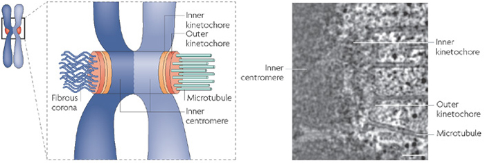
Vertebrate kinetochore ultrastructure.
(A) Microtubules insert into a protein complex called the kinetochore; the kinetochore is an assemblage of proteins on the centromere. (B) An electron micrograph of a human kinetochore; image courtesy of Y. Dong and B. McEwen, State University of New York at Albany, USA. Scale bar, 100 nanometers (nm).
This image is linked to the following Scitable pages:

The five phases of mitosis and cell division tightly coordinate the movements of hundreds of proteins. How did early biologists unravel this complex dance of chromosomes?

Without centromeres, cells cannot divide properly and the overall process of mitosis fails. Why are these small chromosomal regions so essential to such a major cellular process?


Comments
CloseComments
Please Post Your Comment