Evolution of ideas for spindle organization and force generation.
- Author: T. J. Mitchison
Keywords
Flag Inappropriate
Delete Content

Evolution of ideas for spindle organization and force generation.
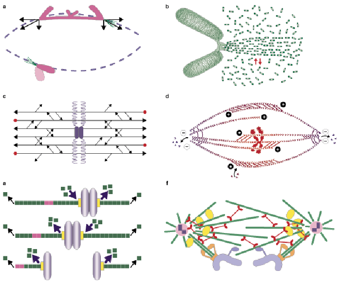
(a) Ostergren's view in the late 1940s. The image represents the spindle in an insect spermatocyte. The forces acting on the chromosomes (arrows) were inferred from chromosome stretching and time-lapse imaging of movement. Ostergren argued that metaphase alignment was due to equalization of pulling forces when the chromosome was centered on the metaphase plate. (b) Inoue's view in the mid-1960s. Here, protein-assembly dynamics are central to force production, but the structural details are unclear. Later, Inoue concluded that tubulin must be polymerized at kinetochores during metaphase, and depolymerized near the poles. (c) The model proposed by McIntosh et al. in 1969, which explicitly invokes microtubule polarity and motor proteins. Horizontal arrows indicate microtubules and their polarity; short diagonal arrows represent putative mechanochemical enzymes. The predicted microtubule polarity is incorrect, but this model was important in highlighting the importance of polarity and in stimulating the search for motor proteins involved in mitosis. (d) Margolis and Wilson's 1981 treadmilling model. Arrows show sites of polymerization at kinetochores and free microtubule plus ends, and depolymerization at poles; they also indicate microtubule polarity. This model combined knowledge of tubulin biochemistry and spindle-microtubule polarity (polarity is indicated with plus and minus signs) and results indicating poleward flux. It predicted poleward flux and postulated functions for dynamics and motors that are still relevant. Whether treadmilling driven by GTP hydrolysis on tubulin has a role in poleward flux remains to be determined. (e) Current model for polymerization dynamics of kinetochore microtubules, showing the role of dynamics at kinetochores and poles in vertebrate tissue-culture cells. The red mark represents a fiduciary mark. Depolymerization at kinetochores, which was discovered by marking experiments, now has a key role in chromosome movement. Poleward flux, which has now been directly observed, is also important. (f) Current motor-centric model that emphasizes the role of Eg5 (red) and dynein (yellow) in organizing the spindle. Kinetochores (orange) are shown moving poleward through the action of a motor (also orange) that may be dynein or an unknown motor protein. Another motor possibly involved in chromosome movement is attached to chromosome arms (blue). The minus ends of spindle microtubules are shown detached from the centrosomes to allow flux.


Comments
CloseComments
Please Post Your Comment