Developmental alterations in the Arabidopsis double chmp1a;chmp1b mutant
Keywords
Flag Inappropriate
Delete Content
Developmental alterations in the Arabidopsis double chmp1a;chmp1b mutant
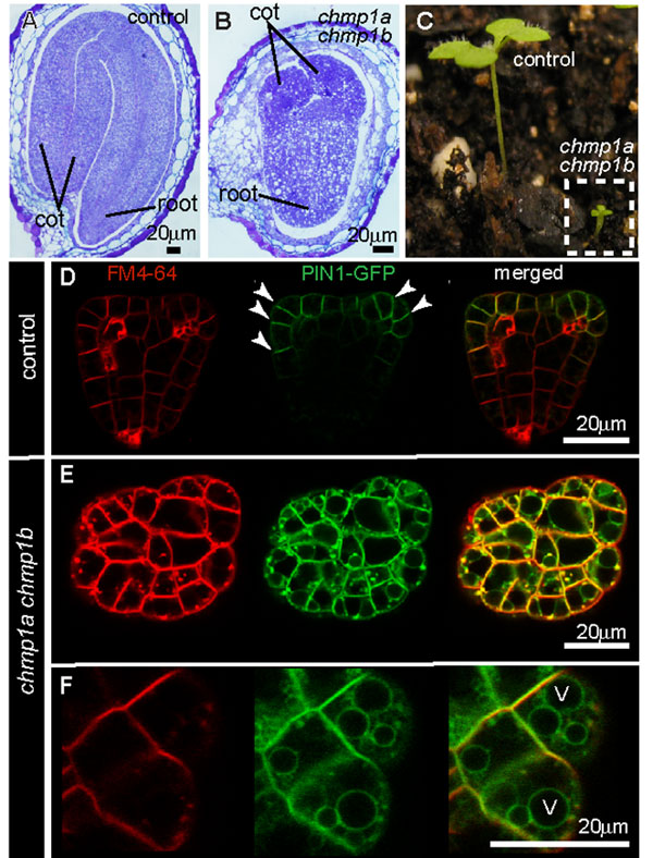
(A) and (B) comparison between control and double chmp1a;chmp1b mutant embryos within their seeds. (C) Seedlings of control and chmp1a;chmp1b plants grown side by side in soil. (D-F) Images obtained with a laser scanning confocal microscope showing the distribution of PIN1 in control (D) and chmp1a;chmp1b mutant embryos (E, F). The PIN1 protein was fused to a green fluorescent protein (GFP) by molecular biology approaches, and now the green fluorescence of the PIN1-GFP protein can be detected in embryo cells. Note that whereas PIN1-GFP signal is detected mostly at the plasma membrane of control cells (arrowheads), PIN1-GFP is also detected in rings that represent the vacuolar membrane (V) in chmp1a;chmp1b mutant cells (F). The fluorescent dye FM4-64 binds the plasma membrane and is internalized by endocytosis, allowing researchers to follow the fate of endocytosed material in living cells.
This image is linked to the following Scitable pages:

An entire fleet of cellular vesicles called endosomes shuttle proteins from their origin to their destination. Do plants and animals have similar shuttling systems?


Comments
CloseComments
Please Post Your Comment