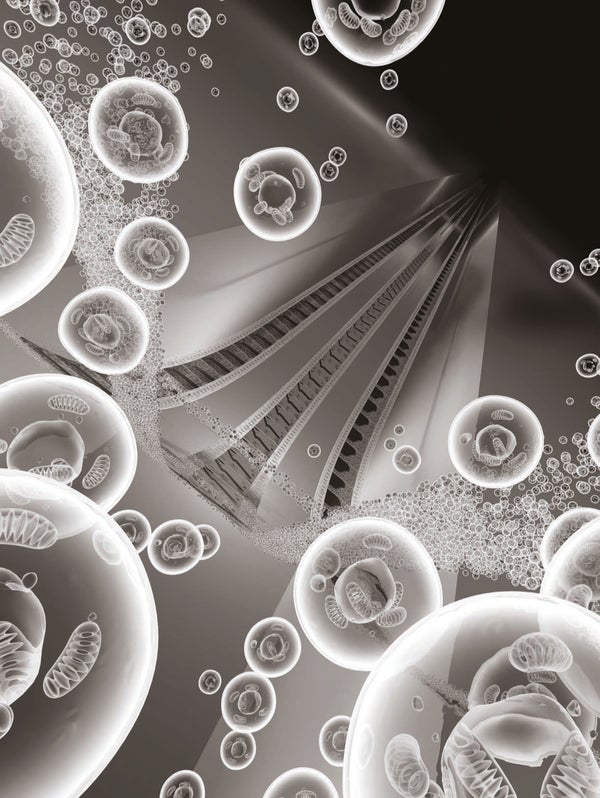
The human eye is limited in its vision. We cannot see objects much thinner than a human hair (a fraction of a millimeter) or resolve motions quicker than a blink (a tenth of a second). Advances in optics and microscopy over the past millennium have, of course, let us peer far beyond the limits of the naked eye, to view exquisite images such as a micrograph of a virus or a stroboscopic photograph of a bullet at the millisecond it punched through a lightbulb. But if we were shown a movie depicting atoms jiggling around, until recently we could be reasonably sure we were looking at a cartoon, an artist's impression or a simulation of some sort.
In the past 10 years my research group at the California Institute of Technology has developed a new form of imaging, unveiling motions that occur at the size scale of atoms and over time intervals as short as a femtosecond (a million billionth of a second). Because the technique enables imaging in both space and time and is based on the venerable electron microscope, I dubbed it four-dimensional (4-D) electron microscopy. We have used it to visualize phenomena such as the vibration of cantilevers a few billionths of a meter wide, the motion of sheets of carbon atoms in graphite vibrating like a drum after being “struck” by a laser pulse, and the transformation of matter from one state to another. We have also imaged individual proteins and cells.
Four-dimensional electron microscopy promises to answer questions in fields ranging from materials science to biology: how to understand the behavior of materials from the bottom up, from the atomic to macroscopic scale; how nanoscale or microscale machines (NEMS and MEMS) function; and how proteins or assemblies of biological molecules fold and become organized into larger structures, a vital process in the functioning of all living cells. Four-dimensional microscopy can also reveal the atomic arrangements of nanoscale structures (which determine the properties of new nanomaterials), and, potentially, track electrons moving around in atoms and molecules on the timescale of attoseconds (a billion billionth of a second). Along with the advances in basic science, the potential applications are wide-ranging, including the design of nanomachines and new kinds of medicines.
On supporting science journalism
If you're enjoying this article, consider supporting our award-winning journalism by subscribing. By purchasing a subscription you are helping to ensure the future of impactful stories about the discoveries and ideas shaping our world today.
Cats and Atoms in Motion
Although 4-D microscopy is a cutting-edge technique that relies on advanced lasers and concepts from quantum physics, many of its principles can be understood by considering how scientists developed stop-motion photography more than a century ago. In particular, in the 1890s, Étienne-Jules Marey, a professor at the Collège de France, studied fast motions by placing a rotating disk with slits in it between the moving object and a photographic plate or strip, producing a series of exposures similar to modern motion picture filming.
Among other studies, Marey investigated how a falling cat rights itself so that it lands on its feet. With nothing but air to push on, how did cats instinctively perform this acrobatic feat without violating Newton's laws of motion? The fall and the flurry of legs took less than a second—too fast for the unaided eye to see precisely what happened. Marey's stop-motion snapshots provided the answer, which involves twisting the hindquarters and forequarters in opposite directions with legs extended and retracted. High divers, dancers and astronauts learn similar motions to turn themselves.
Another approach, stroboscopic photography, relied on short light flashes to capture events occurring on much shorter timescales than is possible with mechanical shutters. The flashes make an object moving in the dark momentarily visible to a detector such as an observer's eye or a photographic plate. In the mid-20th century Harold Edgerton of the Massachusetts Institute of Technology greatly advanced stroboscopic photography by developing electronics that could produce reliable, repetitive, microsecond flashes of light.
The falling-cat experiment requires shutter times or stroboscopic flashes short enough for the photographs to show the animal clearly despite its motion. Suppose the cat has righted itself half a second after being released. At that instant the cat will be falling at five meters per second, so by using one-millisecond flashes we will ensure that the cat falls no more than five millimeters during each exposure so that the image of the cat will be only slightly blurred by its motion. To slice the acrobatics into 10 snapshots, the photographs must be taken every 50 milliseconds.
If we wish to observe the behavior of a molecule instead of a feline, how fast must our stroboscopic flashes be? Many changes in molecular or material structure involve atoms moving a few angstroms (one angstrom equals 10
−10 meter). To map out such motion requires a spatial resolution of less than one angstrom. Atoms often move at speeds of about one kilometer per second in these transformations, requiring stroboscopic flashes no longer than 10 femtoseconds to observe them with better than 0.1-angstrom definition. As long ago as the 1980s researchers used femtosecond laser pulses to time chemical processes involving moving atoms, but without imaging the positions of the atoms in space—the wavelength of the light is hundreds of times longer than the spacing between atoms in molecules or materials [see “The Birth of Molecules,” by Ahmed H. Zewail; Scientific American, December 1990].
Accelerated electrons have long produced images at atomic scales—as in electron microscopes—but only with targets fixed in place and imaged over time intervals of milliseconds or longer, being limited by the speed of the camera. The atom-scale movies we sought thus required the spatial resolution of an electron microscope but with femtosecond electron pulses to “illuminate” the targets. The illuminating packets of electrons are called probe pulses.
Another issue is clocking of the motion—having a well-defined instant in time when the motion begins. We will not get useful images if all the probe pulses take snapshots before the motion starts or after it finishes. In photographing the cat, the recording begins when the cat is released. For ultrafast recording, a femtosecond initiation pulse called the clocking pulse launches the material or the process to be studied.
Even with probing and clocking under control, the issue of synchronization remains. Here the typical ultrafast experiment drastically departs from the cat analogy. Marey could complete his experiment by dropping one cat once, if everything went according to plan. And it did not matter much if the series of exposures began, say, five, 10 or 17 milliseconds after the cat's release. Ultrafast microscopy, however, may probe millions of atoms or molecules for each clocking pulse or may build up images by repeating an experiment thousands of times. Imagine if Marey had been restricted to capturing only a narrow vertical strip of the field of view with each cat drop. To build up the series of full snapshots of the falling cat, he would have had to repeat the experiment many times, recording along a slightly different vertical strip each time. For the various strips to combine sensibly and form a meaningful whole image, he would need to prepare the cat in the same starting configuration for each drop and carefully synchronize the release with the shutter openings in the same way each time. (The technique would also rely on the cat moving in the same fashion every time. I suspect molecules are more reliable than cats in that respect.)
The starting configurations must be accurate to a small fraction of the cat's size, and the time synchronization must be accurate to less than the shutter durations. Similarly, in ultrafast imaging of atoms or molecules, the launch configuration must be defined to subangstrom resolution, and the relative timing of clocking and probe pulses must be of femtosecond precision. The timing of probe pulses relative to the clocking is accomplished by sending either of these pulses along a path with an adjustable length. For a pulse traveling at the speed of light, setting the path length to an accuracy of one micron corresponds to setting the relative timing with 3.3-femtosecond accuracy.
A further major and fundamental problem remained to be overcome before we could make movies with electrons. Unlike photons, electrons are charged and repel one another. Crowding a lot of them into a pulse spoils both the temporal and spatial resolutions because the electrons' mutual repulsion blows the pulse apart. In the 1980s Oleg Bostanjoglo of the Technical University of Berlin did achieve imaging using pulses having as few as 100 million electrons, but the resolutions were no better than nanoseconds and microns (later significantly improved to the submicron level by researchers at Lawrence Livermore National Laboratory).
My group attacked this challenge by developing single-electron imaging, which built on our earlier work with ultrafast electron diffraction. Each probe pulse contains a single electron and thus provides only a single “speck of light” in the final movie. Yet thanks to each pulse's careful timing and another property known as the coherence of the pulse, the many specks add up to form a useful image of the object. A similar feat is sometimes exhibited as one of the characteristic oddities of quantum mechanics: electrons pass through two slits one at a time, each one contributing a single speck at some random location on a detection screen. Yet all the specks add up to form predictable patterns of light and darkness characteristic of interfering waves.
Single-electron imaging was the key to 4-D ultrafast electron microscopy (UEM). We could now make movies of molecules and materials as they responded to various situations, like so many startled cats twisting in the air.
Deciphering Nanomatter
One of our first targets was graphite, the “lead” material in pencils. We chose graphite in part because it is an unusual material, with applications in environments as extreme as those in nuclear reactor cores, and because it has close relatives that are just as remarkable. Graphite consists of carbon atoms arranged in a hexagonal pattern to form sheets reminiscent of chicken wire. Relatively weak bonds hold the sheets together in a stack. Writing with an ordinary pencil relies on pieces of the graphite sloughing off and adhering to the paper. The pencil marks include tiny quantities of the strongest material known to science—graphene, which consists of isolated single sheets of carbon atoms. Researchers are studying graphene vigorously for a variety of electronics applications. Furthermore, when soft graphite is subjected to extreme pressure, its atoms rearrange to form diamond, one of the hardest known substances.
To study graphite's response to mechanical shocks, we took nanoscale crystals of the substance—some only nanometers thick, or a few sheets of atoms—and struck them with intense femtosecond laser pulses, which served as the clocking pulses for our microscope. Each laser pulse pushed the graphite's layers of atoms momentarily closer together, setting them oscillating up and down [see box on opposite page]. Our electron microscope sent its electrons through these oscillating graphite layers to produce two kinds of picture: a real-space image (much like a photograph of the graphite surface) or a diffraction pattern, which is a regular array of spots whose precise configuration provides information about the arrangement and separations of atoms in the graphite lattice. In particular, we could track the layers oscillating up and down by the movements of the spots in the diffraction pattern. The oscillations had frequencies of about 10 to 100 gigahertz (10
10 to 1011 cycles per second). No imaging experiment had previously observed such high-frequency resonances unfolding over time.
From our measurements we determined the elasticity of graphite perpendicular to the planes of atoms—how the material responds to compressing or stretching forces acting in that direction. Imagine that the graphite crystal is a stack of rigid metal plates connected by springs and that the laser pulse is a large sledgehammer striking the top plate. We measured the properties of the springs.
The metal-plate analogy is reasonable as long as our “camera” is zoomed in very close. If the camera figuratively “pulls back,” however, more of the tiny graphite crystal comes into view. Now the hammer is striking one region of the top metal sheet, and it becomes apparent that the sheets are flexing, with the compression and expansion propagating out from the impact point in waves.
When we pull back the camera even farther and take images more slowly, yet another kind of dynamics comes into view. Now we see how the laser pulse sets the entire nanoscopically thin crystal oscillating, like a drumhead hit by a drumstick. We saw that in the first few microseconds after the laser pulse hit, the crystal's motion appeared chaotic, but as time went on the entire crystal settled down into a well-defined resonant oscillation—it drummed!
For these oscillations, the material property that sets the resonance frequency is the elasticity of the graphite planes—their response to being stretched or compressed in the plane. We found that the graphite is much more resistant to being deformed in the planes of carbon atoms than it is to having those planes pulled apart or pushed together. The results can be explained by considering that the chemical bonds joining the carbon atoms in each hexagonal layer are much stronger than the bonds linking adjacent planes to one another.
Although studies of bulk samples of graphite produce similar data about graphite's elasticity, the information we obtained tells us much more. It addresses questions of two types that are fundamental to our understanding of how materials behave at the nanoscale: first, at what length scale does the description of a substance in terms of a continuum material with properties such as elasticity break down? Second, can we extrapolate from the behavior at atomic scales of length and time to reproduce the known macroscopic properties of a material? With graphite, we found that even quite nanoscopic samples (only a few dozen atomic layers thick) behave surprisingly like the bulk material. Would this description still be valid near the graphene limit?
The movies of graphite I have described thus far all relied on collisions of our probe electrons with the sample in which they lose no energy—like rubber balls bouncing off something hard. Sometimes, however, a probe electron may lose energy, by exciting an electron in a carbon atom. The amount of energy lost depends on the kind of bond in which the atom's electron was involved. A very old technique called electron energy loss spectroscopy can measure such losses; the energy spectra obtained provide information about the bonding in a material and the chemical elements that compose it. Using this method with our ultrafast electron microscope, we showed that during the compression phase, the bonding inside the graphite shifted toward the kind of bond that is characteristic of diamond. In the expansion phase, the bonding of the surface atoms shifted toward that of graphene. Conventional electron energy loss spectroscopy is far too slow to observe these changes.
From Cantilevers to Cells
My group has now carried out four-dimensional microscopy on a number of materials in addition to graphite. In iron, we made diffraction images to follow the crystal structure changing from what is called body-centered cubic to face-centered cubic, a process that occurs in many industrial applications at high temperatures, including production of steel. We saw two dynamic processes unfold when we heated the iron from room temperature to nearly 1,500 kelvins in about a nanosecond. First, specks of the face-centered phase developed, or nucleated, at locations in the crystal relatively slowly—on a nanosecond timescale—out of the incoherent motions of iron atoms. Second, these regions of the new phase grew at the speed of sound, meaning that the process took only picoseconds (10
−12 second) to encompass the hot iron. This rapidly spreading transformation involves numerous atoms being displaced in a coordinated fashion, a curious kind of “emergence” of a large-scale change in the crystal from the innumerable underlying nanoscopic motions. Understanding of this phenomenon might lead to better ways to handle iron and steel (and many other materials) in industrial processes.
One of the most powerful applications of 4-D ultrafast electron microscopy is seeing nanosystems and microsystems as they function in real time. For instance, we imaged the resonant oscillations of nanoscopic cantilevers, which had not been accomplished before for such high-frequency motions. From our results we determined a range of quantities that describe the cantilevers' material properties and their motion, and we saw that they functioned coherently for nearly 10
11 oscillations. Researchers can use such data to test the theoretical models that guide design of microelectromechanical and nanoelectromechanical systems, which in turn may lead to new kinds of such devices or new uses for them.
Four-dimensional imaging with ultrafast electron microscopy also has potential biological applications. To fully understand how the body functions, investigators need to know not only the structures of the various proteins and other molecular and cellular structures involved but also their dynamics—how a protein folds, how it selectively recognizes other molecules, what role the water around it plays, and so on. Some biological functions involve ultrafast steps. For instance, our vision and photosynthesis in plants both rely on photons of light triggering femtosecond-scale processes. Although many proteins function, and malfunction, on timescales much longer than femtoseconds, the atomic and molecular motions in the initial femtoseconds can determine whether these macromolecules ultimately fold properly into a useful structure or into one that, say, causes Alzheimer's disease.
One study on protein folding illustrates the kind of techniques needed and the results that are possible. My colleagues and I investigated how quickly a short length of protein would fold into one turn of a helix by heating the water in which the protein was immersed—a so-called ultrafast temperature jump. (Helices occur in innumerable proteins.) We found that short helices formed more than 1,000 times faster than researchers have thought—arising in hundreds of picoseconds to a few nanoseconds rather than the microseconds commonly believed. Knowing that such rapid folding occurs may lead to new understanding of biochemical processes, including those involved in diseases.
Biological imaging with our 4-D ultrafast technology often relies on a well-established technique called cryoelectron microscopy, in which a sample in water is plunged quickly into liquid ethane (which boils at −89 degrees Celsius). The water freezes into a glassy solid that does not diffract electrons and spoil imaging (and the sample itself!) as ordinary ice crystals do. We have obtained images of bacterial cells and protein crystals in this way. In the future we hope to watch proteins embedded in such vitreous water fold and unfold: a clocking pulse will boost the temperature enough to melt a tiny droplet of the water around the protein, which will unfold and then promptly refold. When the water cools and refreezes, it renders the molecule ready for another clocking pulse. The same approach could allow us to visualize the dynamics of bacterial flagella and of the fatty acid bilayers that make up cell membranes. As with our graphite studies, ultrafast electron energy loss spectroscopy should let us map changes in bonding. Capturing the image before the biosystem moves or disintegrates should provide sharper images than currently possible in cryomicroscopy.
Variants of ultrafast electron microscopy might well push below the nanoscale in structural dynamics studies and below a femtosecond in the imaging of the electron distribution in matter. Very recently, my Caltech group demonstrated two new techniques. In one, convergent-beam UEM, the electron pulse is focused and probes only a single nanoscopic site in a specimen. The other, near-field UEM, enables imaging of the evanescent electromagnetic waves (“plasmons”) created in nanoscopic structures by an intense laser pulse—a phenomenon that underlies an exciting new technology known as plasmonics [see “The Promise of Plasmonics,” by Harry A. Atwater; Scientific American, April 2007]. This technique has produced images of bacterial cell membranes and protein vesicles with femtosecond- and nanometer-scale resolution.
In recent years Ferenc Krausz of Ludwig Maximilian University of Munich, Paul Corkum of the University of Ottawa and others have opened up the attosecond regime to optical (light-based) studies using extremely short laser pulses. At Caltech, we have proposed several ultrafast electron microscopy schemes for attosecond-scale electron-based imaging, and we are now pursuing the experimental realization in collaboration with Herman Batelaan of the University of Nebraska–Lincoln.
The electron microscope is extraordinarily powerful and versatile. It can operate in three distinct domains: real-space images, diffraction patterns and energy spectra. It is used in applications ranging from materials and mineralogy to nanotechnology and biology, elucidating static structures in tremendous detail. By integrating the fourth dimension, we are turning still pictures into the movies needed to watch matter's behavior—from atoms to cells—unfolding in time.