
Thank you for visiting nature.com. You are using a browser version with limited support for CSS. To obtain the best experience, we recommend you use a more up to date browser (or turn off compatibility mode in Internet Explorer). In the meantime, to ensure continued support, we are displaying the site without styles and JavaScript.

Volume 81 Issue 6, June 2017

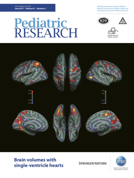
Watson et al. obtained magnetic resonance images of the brains of 10- to 19-year-olds who had undergone the Fontan procedure for single-ventricle defects and compared them with those of healthy controls. They found significant and widespread reduction in regional gray matter. Several surgery-related issues were associated with reduction in volume, suggesting potentially modifiable risk factors. See the article on page 881.

Editor's Focus

Top of page ⤴

Commentary

Top of page ⤴

Population Study

Top of page ⤴

Clinical Investigation

Top of page ⤴

Translational Investigation

Top of page ⤴

Basic Science Investigation

Top of page ⤴

Search

Quick links