Thank you for visiting nature.com. You are using a browser version with limited support for CSS. To obtain
the best experience, we recommend you use a more up to date browser (or turn off compatibility mode in
Internet Explorer). In the meantime, to ensure continued support, we are displaying the site without styles
and JavaScript.
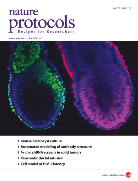
Peri-implantation embryogenesis is largely unexplored and probably the most enigmatic period of mouse development. The protocol by Bedzhov et al. describes how to support murine embryonic development beyond the blastocyst stage in vitro allowing direct observation of blastocyst to egg cylinder transition. The egg cylinder shown on the cover was recovered at E6.5. Oct4 (red) marks the epiblast. Eomes (green) is expressed in the posterior EPI, extraembryonic ectoderm and visceral endoderm. DAPI (blue) marks the nuclei.
Pancreatic ductal infusion enables the delivery of dyes or viruses directly to specific pancreatic cell types, permitting their manipulation. This manipulation is helpful for the study of diabetes, pancreatitis and pancreatic cancer.
Murine inner-medullary collecting duct cells grown in a matrigel mixture grow into spheroids with apicobasal polarity to model renal disease and test the functional relevance of new gene mutations.
The morphogenetic and signaling events during blastocyst to egg cylinder transition at the peri-implantation stages are largely unexplored. This protocol supports development of mouse embryos beyond the blastocyst stage in vitro.
This protocol describes how to maintain the gastrointestinal nematode Heligmosomoides polygyrus bakeri and generate excretory-secretory products for identification, cloning, and immunological characterization of the modulatory molecules.
This protocol recreates the in vivo condition of latently infected, resting CD4+ T lymphocytes and can be used for drug screening, studying CTL responses to HIV-1, comparing viral alleles, or growth of cells from HIV-1–infected individuals.
Predicting the structure of antibodies on the basis of their sequences is a key objective in medical and biotechnology research. Marcatili et al. describe the use of their online system PIGS for the automated modeling of the 3D structure of antibodies.
This protocol describes how to rapidly anchor pipettes to the head of a rat, enabling reliable whole-cell recording of neurons from freely moving rats and facilitating studies of neuronal integration and plasticity in identified cells during natural behaviors.
Biochemical methods have typically been underused in Drosophila research owing to technical challenges. Here Depner et al. describe a simple CNS fractionation method that yields ∼4 mg of synaptic membrane protein per 1 g of adult fly heads.
This protocol describes how to prepare mouse, rat, human and porcine pancreas tissue slices and how to use them to investigate cellular mechanisms underlying the function, pathology and interaction of endocrine and exocrine components of the pancreas.
This protocol describes the preparation and application of a polymer-based nanosensor that combines the two pH-sensitive fluorophores fluorescein and Oregon Green for imaging intracellular pH (pH 3.1–7.0).
In this Protocol, data obtained from optical projection tomography and confocal microscopy is analyzed with Tree Surveyor and Imaris to obtain information about branching morphogenesis. Mouse kidneys are used as an example, but the method is applicable to other branched organs.
Gargiulo et al. describe the use of shRNA libraries for functional screens in orthotopic mouse tumor models, using glioma and lung cancer as examples. Guidelines are included for adapting the method to other tumor models or technologies (e.g. CRISPR).