
Volume 7 Issue 8, August 2012
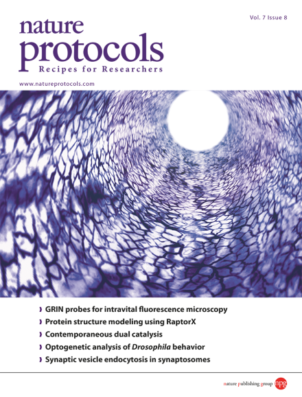
A rendering of the vasculature of the colon of a mouse. The image was produced from the 3D data set acquired in vivo using a small-diameter graded-index (GRIN) probe integrated into a confocal fluorescence microscope. (The diameter of the lumen is ∼1.5 mm.) Image taken from the protocol by Kim et al. 10.1038/nprot.2012.078.
Protocol
-
Advertisement
