
Volume 15 Issue 5, 1 May 2020
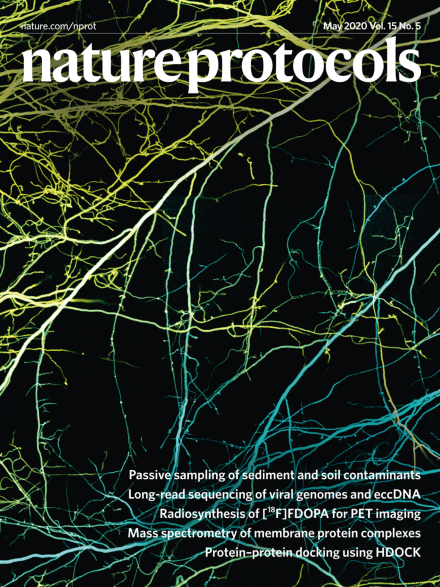
Confocal image of Neurospora crassa hyphae
Neurospora crassa hyphae expressing CFP (blue) or YFP (yellow). Green are fused hyphae with mixed nuclei in a common cytoplasm. Maximum intensity projection of 3 × 3 tiled z-stack of confocal images. Image is 2.0 × 2.7 mm.
See: Jonkman et al.
Image: Work was conducted at The Rockefeller University. Sample prepared by Gregory Jedd, Temasek Life Sciences Laboratory, during postdoctoral research; image acquired by Alison North. Cover Design: Erin Dewalt
Review Articles
-
Advertisement
