
Volume 11 Issue 6, June 2016
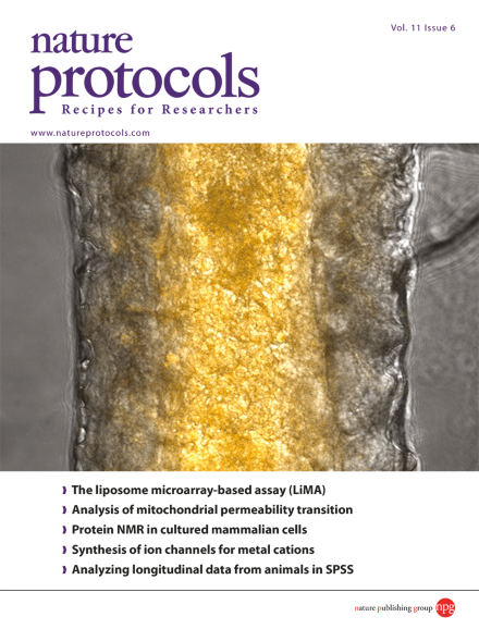
Chen et al. describe a methodology for the preparation of a rhodamine-based HOCl probe, R19S, and its use for the detection of HOCl in living cells and organisms. Shown is a confocal image of a Drosophila intestine following enteric infection with a Drosophila pathogen, Erwinia carotovora carotovora. Pathogen-induced HOCl molecules in the gut lumen are labeled yellow-orange. Image taken from the protocol by Chen et al.
Protocol
-
Advertisement
