Thank you for visiting nature.com. You are using a browser version with limited support for CSS. To obtain
the best experience, we recommend you use a more up to date browser (or turn off compatibility mode in
Internet Explorer). In the meantime, to ensure continued support, we are displaying the site without styles
and JavaScript.
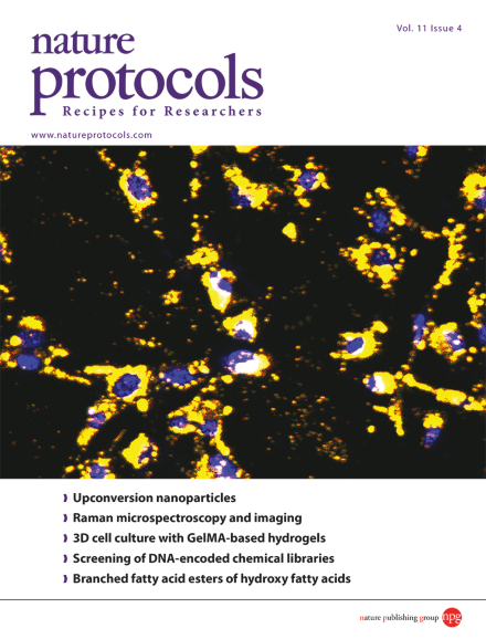
Upconversion nanoparticles taken up by cultured melanoma cells emit yellow luminescence under 980-nm laser excitation (blue fluorescence from 4',6-diamidino-2-phenylindole (DAPI) counterstaining indicates the cell nucleus). The upconversion luminescence activates photosensitizers encapsulated within the mesoporous silica coating of the nanoparticles, producing toxic reactive oxygen species that induce death of the cancer cells. Taken from the protocol by Gnanasammandhan et al. doi: 10.1038/nprot.2016.035. Cover design by Jamel Wooten.
Interferometric scattering microscopy (iSCAT) enables ultrasensitive label-free imaging and high-speed single-particle tracking. This protocol describes how to construct an iSCAT microscope with single-molecule TIRF capabilities.
This protocol describes how to set up and use the autopatcher, a robot that automatically obtains intracellular neural recordings from intact mammalian mouse brains.
Annexin A5 and TO-PRO-3 (a nucleic acid–binding dye that stains early apoptotic and necrotic cells differentially) are used to distinguish six types of particles in a sample, including apoptotic bodies and cells at various stages of cell death.
Raman microspectroscopy is useful for the analysis of biological samples, because chemical and structural information can be obtained without using labels. This protocol brings together practical guidelines from expert research groups.
Upconversion nanoparticles (UCNs) have the extraordinary ability to emit light with UV-visible wavelengths on illumination in the near IR region. This protocol describes the preparation of UCNs with potential therapeutic applications.
This protocol from Hassan et al. describes a microfluidic chip that uses an immunocapture chamber to count CD4 and CD8 cells in whole blood for HIV/AIDS diagnostics. The chip can be adapted for different cell types and research applications.
This protocol describes how to make semisynthetic gelatin methacryloyl (GelMA)-based hydrogels for use in 3D cell culture models for cancer research, stem cell research and tissue engineering.
Branched fatty acid esters of hydroxy fatty acids (FAHFAs) are a recently discovered class of biological lipids. This protocol describes their extraction from serum and tissue samples, followed by enrichment and analysis by LC-MS.
This protocol describes an approach for screening DNA-encoded chemical libraries (DECLs) to identify molecules that bind to proteins of interest. After isolating binding library members, DNA barcodes are amplified and identified by high-throughput sequencing.
Many solid tumors contain an aggressive hypoxic region that is difficult to treat. This protocol describes how to prepare bioreductive prodrugs that are biologically inactive until they are converted to an active drug by enzymatic reduction in hypoxia.
This protocol describes a data-independent acquisition workflow for label-free quantitative proteomics that integrates ion mobility separation and applies drift time–specific collision energies to improve precursor fragmentation efficiency.
Human NET-seq enables DNA strand–specific mapping of RNA polymerase (RNAP) activity at single-nucleotide resolution. A cell fractionation approach is used to isolate transcribing RNAP and associated RNAs, avoiding immunoprecipitation or RNA labeling.