Thank you for visiting nature.com. You are using a browser version with limited support for CSS. To obtain
the best experience, we recommend you use a more up to date browser (or turn off compatibility mode in
Internet Explorer). In the meantime, to ensure continued support, we are displaying the site without styles
and JavaScript.
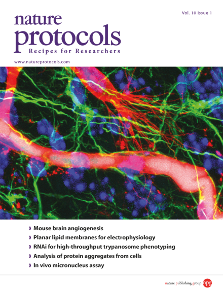
The protocol by Wälchli et al. describes how to monitor newly forming, and established, functional blood vessels in the postnatal mouse brain. An isolectin B4positive (IB4+, red) endothelial tip cell of a sprouting blood vessel extends multiple filopodia opposite to an Evans blue–positive (EB+, cyan)/IB4+ functional, perfused blood vessel in an 8-day-old mouse brain cortex. Cells are stained so that IB4+ blood vessel endothelial cells are red, GFAP+ astrocytes and GFAP+ radial glia are green and DAPI+ cell nuclei are blue.
A challenge for metabolomics analysis is ensuring that sample preparation does not alter the cells' metabolic state. This protocol describes LC/MS analysis of core metabolites from adherent mammalian cells after rapid wash and quenching steps.
This protocol describes a DNA-repair assay that measures UDS, RRS, and cell sensitivity after the incorporation of EdU/EU and detection by click chemistry. It can be used for diagnosing DNA repair deficiency disorders and screening anti-cancer drugs.
This protocol describes how to microinject a DNA plasmid-amphiphilic molecule mix into the neocortex. Subsequent delivery of electric pulses gives rapid, focal, and efficient expression of genes in postmitotic neurons.
Bacterial growth can be monitored by following the incorporation of fluorescent D-amino acids (FDAA) into the peptidoglycan cell walls. This protocol covers the synthesis of FDAAs and their use in monitoring spatiotemporal bacterial growth.
This protocol uses intravascular Evans blue labeling and Isolectin-B4 endothelial tip cell staining combined with antibody-based protein detection to monitor new blood vessel formation from tip cell selection to functional, perfused blood vessels.
Biochemical characterization of mechanosensitive proteins is achieved in a protocol for forming actomyosin cables between protein-coated discs on a micropatterned glass coverslip. Interaction with a fluorescently labeled ligand is analyzed by microscopy.
This protocol describes sortase A-mediated site-specific bioconjugation of single-chain antibodies to iron oxide particles for magnetic resonance imaging and to layer-by-layer capsules for drug delivery.
This protocol describes RIT-seq, an approach for performing genome-scale RNAi library screens in bloodstream-form African trypanosomes, a family of parasites that also serves as a model for studies on aspects of eukaryotic biology and evolution.
Intracellular protein aggregation is a hallmark of different pathological conditions. Hollow fiber flow field-flow fractionation, coupled with multiangle light scattering, can be used to separate and biophysically characterize cellular aggregates.
This protocol uses a highly sensitive and reproducible antibody-based capillary isoelectric focusing approach to establish protein phosphorylation status from nanogram quantities of protein cell lysate from cell line or primary stem cell material.
A platform for isolating low-abundance protein complexes from Arabidopsis seedlings and cell cultures is described. Its power resides in an improved TAP tag combined with ultrasensitive MS and filtering against a list of nonspecific proteins.
Electrophysiology experiments on reconstituted membranes provide functional information on membrane proteins. Protocols for Montal-Mueller and Mueller-Rudin bilayers, or for formation via giant liposomes are presented and compared.
This protocol describes how to immobilize living cells into polydimethylsiloxane stamps, generating an array of living cells that can undergo atomic force microscopy, with no chemical or physical denaturation.
This protocol describes a sensitive, robust, high-throughput method for quantifying the formation of micronuclei in mouse erythrocytes. It detects low levels of genome instability and can be used to identify novel genome maintenance genes and models.