Volume 19
-
No. 4 April 2024
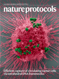
Tumor cell captured via magnetic beads functionalized with tetrahedral DNA frameworksScanning electron micrograph of streptavidin-labeled magnetic beads (green) bound to biotin-labeled tetrahedral DNA frameworks anchored with aptamers to a HepG2 cell (pink). See Chen, Y. et al.
-
No. 3 March 2024
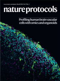
Studying human brain vascular cells with single-cell transcriptomics and organoidsHuman brain vascular cells, including endothelial and mural cells, can be purified with FACS and used in many downstream applications, including cell culture, transcriptomics and organoid transplants. This image shows vascular cells labeled with GFP and transplanted on top of induced pluripotent stem cell (iPSC)-derived cortical organoids. See Crouch et al. p603
-
No. 2 February 2024
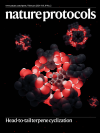
Terpene cyclization inside the resorcin[4]arene capsuleHexameric capsules that self-assemble from resorcin[4]arene are shown in this rendered image. Luminous oxygen and hydrogen atoms highlight the hydrogen bond network holding the assemblies together. In the foreground, the capsule is open, revealing its content: a cyclized presilphiperfolanol molecule. See Cornu et al.
-
No. 1 January 2024
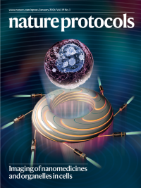
Label-free X-ray microscopy of nanomedicines and organelles in intact single cells at nanometer resolution using synchrotron radiationA composite image of a single cell visualized in 3D and a synchrotron radiation facility. The method uses X-rays generated via synchrotron radiation and enables the subcellular localization of nanomedicines in single cells, at nanometer resolution, as a robust approach to characterize interactions between nanomaterials and cells. See Cao et al.