Volume 32 Issue 5, May 2007
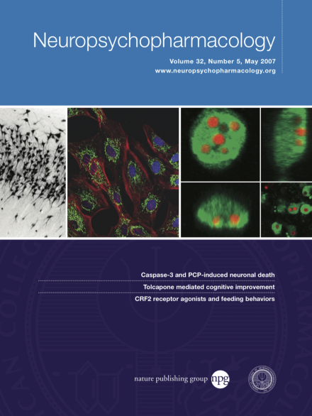
Confocal images (xy, yz and xz planes) of phencyclidine-induced apoptosis in the hippocampus of a PN7 rat nine hrs after administration (10 mg/kg). Caspase-3 immunoreactivity (green) is largely confined to the cytoplasm surrounding a multifragmented nucleus (red) in which cleaved DNA is visualized using terminal d-UTP nick-end labeling (TUNEL). The lower right panel shows similar caspase-3 and TUNEL staining of layer II–IV parietal cortex neurons 12 hrs after addition of 3 μM PCP to cultured cortical slices. Refer to figures 4f and 7f for additional details. [Neuropsychopharmacology, 2007, 32, 1178–1194]