Volume 13
-
No. 12 December 2016
Combining literature-curated resources (lenses) in the Omnipath tool (glasses) gives a clearer view of signaling pathways and improves prior knowledge for downstream data analysis. Artwork by S. Phillips, EMBL-EBI; idea by J. Wirbel, RWTH Aachen. Correspondence p966
-
No. 11 November 2016
Confocal image of a four-day-old zebrafish embryo (10× objective) stained for markers of basal stem cells (green), surface epithelial cells (red) and all nuclei (blue). Image acquired by Oscar Ruiz in the laboratory of George Eisenhoffer at MD Anderson Cancer Center. Winner of the 2016 Nikon Small World photomicrography contest (reprinted with permission from O.R. and G.E.; image provided by Nikon).
-
No. 10 October 2016
Pseudocolored mouse brain after tissue clearing with uDISCO. Cover image by Ruiyao Cai and Ali Ertürk; edited by Erin Dewalt.
-
No. 9 September 2016
On the cover: smURFPs are far-red fluorescent proteins derived from a cyanobacterial phycobiliprotein that perform well in a range of molecular imaging applications. Cover by Erik A. Rodriguez (images and artwork) and Erin Dewalt (image editing). Article p763.
-
No. 8 August 2016
Griss et al. shed some light on the dark proteome: by clustering both known and unidentified peptide tandem mass spectra across the entire PRIDE Archive repository, they identified more than 9 million previously unidentified spectra. Cover by Erin Dewalt. Resource p651
-
No. 7 July 2016
Fluorescent proteins spanning the visible spectrum were directly compared to determine their relative performance. This image provides an artistic representation of fluorescent proteins as paint swatches. Cover by Erin Dewalt. Analysis p557
-
No. 6 June 2016
A modified protocol that makes expansion microscopy compatible with conventional antibodies and fluorescent proteins was used to generate super-resolution images of dividing cells. Cover by Aaron Halpern. Brief Communication p485
-
No. 5 May 2016
A microfluidic chip enables automated and highly efficient reprogramming of human somatic cells to induced pluripotent stem cells, which can also be differentiated on-chip into different cell types. Cover by Stefano Giulitti, Camilla Luni and Nicola Elvassore (images and artwork) and Erin Dewalt (image editing). Article p446
-
No. 4 April 2016
Scientists attempt to reconstruct signaling networks from indirect measurements (phosphoproteomic changes upon perturbation), akin to prisoners deciphering projected shadows of reality in Plato's allegory of the cave. Cover by Spencer Phillips, EMBLEBI (artwork); Julio Saez Rodriguez, RWTH Aachen and EMBL-EBI (concept); and Erin Dewalt (image editing). Analysis p310
-
No. 3 March 2016
An artistic interpretation of footprints detected on a beach, by Erin Dewalt.
-
No. 2 February 2016
Reconstruction of the force field around a cell within a collagen matrix. Cover based on a figure from Steinwachs et al. Article p171
-
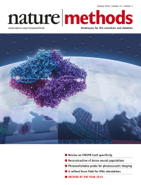
No. 1 January 2016
Single-particle cryo-electron microscopy (cryo-EM) is our choice for Method of the Year 2015 for its newfound ability to solve protein structures at near-atomic resolution. Featured is the 2.2-å cryo-EM structure of β-galactosidase as recently reported by Bartesaghi et al. (Science 348, 1147–1151, 2015). Cover design by Erin Dewalt.Special feature starts on p19.