Thank you for visiting nature.com. You are using a browser version with limited support for CSS. To obtain
the best experience, we recommend you use a more up to date browser (or turn off compatibility mode in
Internet Explorer). In the meantime, to ensure continued support, we are displaying the site without styles
and JavaScript.
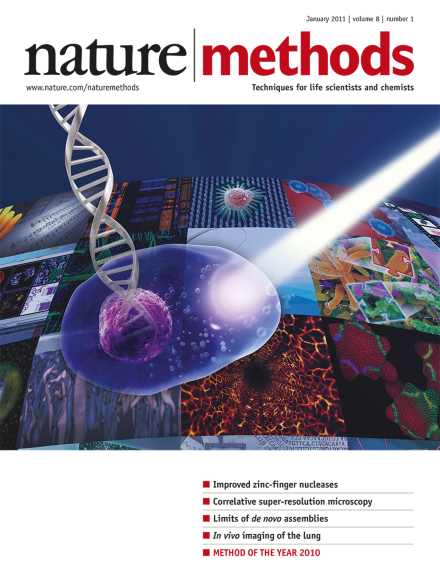
Optogenetics, our pick for Method of the Year 2010, is rapidly becoming a vital tool for neurobiologists, who are applying it to study both basic biology and disease. Cover design by Erin Dewalt. Special feature starts on p19.
With the capacity to control cellular behaviors using light and genetically encoded light-sensitive proteins, optogenetics has opened new doors for experimentation across biological fields.
A team of researchers applied a 'discovery single-particle profiling' experimental strategy to visualize the assembly of the ribosome via time-resolved electron microscopy.
Optogenetics is a technology that allows targeted, fast control of precisely defined events in biological systems as complex as freely moving mammals. By delivering optical control at the speed (millisecond-scale) and with the precision (cell type–specific) required for biological processing, optogenetic approaches have opened new landscapes for the study of biology, both in health and disease.
Optogenetics is routinely used to activate and inactivate genetically defined neuronal populations in vivo. A second optogenetic revolution will occur when spatially distributed and sparse neural assemblies can be precisely manipulated in behaving animals.
Optogenetic modules offer cell biologists unprecedented new ways to poke and prod cells. The combination of these precision perturbative tools with observational tools, such as fluorescent proteins, may dramatically accelerate our ability to understand the inner workings of the cell.
Rhodopsins from microalgae and eubacteria are powerful tools for manipulating the function of neurons and other cells, but these tools still have limitations. We discuss engineering approaches that can help advance optogenetics.
The low cost of short-read sequencing has motivated the development of de novo assemblies from only short-read data; impressively, assemblies for large mammalian genomes are now available. However, this is still a developing field, and these de novo assemblies have many artifacts, as do all de novo assemblies.
Context-dependent assembly (CoDA) of zinc finger nucleases is reported. Starting from an archive of zinc finger modules known to function well together, effective multifinger arrays can be constructed using standard techniques. Also in this issue, Doyon et al. report rational design of nucleases with improved cleavage activity.
A statistical framework for assigning confidence scores for protein-protein interaction data generated via affinity purification–mass spectrometry, called significance analysis of interactome (SAINT) is described.
Identification of residues critical for dimerization of the Fok1 nuclease domain of zinc-finger nucleases permits rational design of enzymes with improved cleavage activity and retained obligate heterodimerization. Also in this issue, Sander et al. report context-dependent assembly (CoDA), a simple method for designing zinc-finger nucleases.
Methods are reported for the combination of fluorescence nanoscopy using either stimulated emission depletion microscopy (STED) or photoactivated localization microscopy (PALM) with electron microscopy, to achieve correlative imaging in which the super-resolved fluorescence signal is placed in the context of cellular ultrastructure.
Derivatizing glycosphingolipids, extracted from bovine brain or human erythrocytes, with a fluorescent tag allows their immobilization on an array which can be probed with glycan binding proteins.
Fast, two-photon intravital imaging of a mechanically stabilized and physiologically intact preparation of the mouse lung is reported. It is used to monitor immune cells in the lung under normal and injured conditions.
Nature Methods' choice of Method of the Year 2010 is optogenetics for its capacity to control cell function with light. A series of articles and a video describe how optogenetics has revolutionized the way experiments are conducted in neuroscience and showcase the potential the method has for the study of many signaling pathways in cell biology. The special feature also discusses how technological development will be needed to expand the possibilities of optogenetics.