Thank you for visiting nature.com. You are using a browser version with limited support for CSS. To obtain
the best experience, we recommend you use a more up to date browser (or turn off compatibility mode in
Internet Explorer). In the meantime, to ensure continued support, we are displaying the site without styles
and JavaScript.
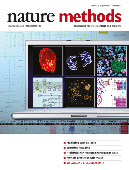
The cover image shows a range of data visualizations currently used by life scientists. Source images come from figures in the Nature Methods supplement "Visualizing biological data" and from Nature Cell Biology and Nature Biotechnology. Cover design by Seán O'Donoghue and Bang wong. Supplement Foreword p193
Getting young researchers into independent positions that encourage risk-taking would benefit science but requires more than targeted individual research grants.
The pioneers of the first generation of widely used optogenetic tools now describe new variants that open further possibilities for studying neural function.
A method to differentially label each sister chromatid in a cell makes it possible to elucidate segregation patterns after mitosis and should help to pinpoint the mechanism behind nonrandom segregation in certain cell types.
A new amyloid-prediction tool, Waltz, offers advantages over previous amyloid-prediction tools for distinguishing 'true' amyloids from amorphous aggregates.
Prospective isolation of defined cell types is a crucial prerequisite for their molecular analysis, but the heterogeneity of populations yielded by current protocols obscures relevant information. New studies now use additional features from time-resolved imaging data for live prospective identification of cells with defined future behavior.
Simple minicircle vectors carrying four reprogramming factors induce pluripotency in adult human adipose stem cells and in neonatal fibroblasts without integration into the genome.
Conventional extracellular electrode recordings are generally limited to monitoring action potentials. But use of extracellular gold microelectrodes with microspines that are engulfed by a neuron generates efficient electrical coupling and allows detection of both action potentials and subthreshold synaptic potentials with a signal-to-noise ratio similar to that of conventional intracellular recordings.
Single-molecule fluorescence resonance energy transfer (smFRET) is applied in live cells and reveals the conformational changes of individual SNARE proteins upon entering a SNARE complex.
Chemically inducible dimerization probes selectively target proteins to the surface of specific organelles or tether organelles to each other, thus allowing precise spatiotemporal analysis of signaling events.
By subdividing a charge-coupled device (CCD) array into subgroups using a digital micromirror device and offsetting exposure times, temporal pixel multiplexing allows simultaneous high-speed and high-resolution imaging using a single CCD. This imaging modality allows 250 Hz microscopic imaging of fast cellular responses with a 10-Hz 1.3 megapixel camera
The fates of cultured neural progenitor cells can be predicted by algorithmic information theory-based computational analysis of time-lapse images of the cells.
A Cre-loxP–based technique allows triggering of heritable coexpression of a fluorescent marker along with any desired transgene, providing a versatile tool for clonal analysis of gene function in the zebrafish.
The use of membrane-tethered toxins to selectively block ion channel function in vivo is demonstrated. The approach is applied to blockade of voltage-gated calcium channels for inhibition of neurotransmission in the mouse.