Thank you for visiting nature.com. You are using a browser version with limited support for CSS. To obtain
the best experience, we recommend you use a more up to date browser (or turn off compatibility mode in
Internet Explorer). In the meantime, to ensure continued support, we are displaying the site without styles
and JavaScript.
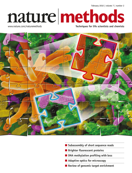
Artistic rendering of an electron micrograph of a methlyamine–enriched metagenomic community from a Lake Washington sediment. Sample was obtained by Marina Kalyuzhnaya and Ludmila Chistoserdova; copyright for the original image, Dennis Kunkel Microscopy, Inc., colorization by Ekaterina Latypova. Cover art by Joseph Hiatt. Brief Communication p119
By sampling a two-dimensional diffraction pattern on a spherical detector, three-dimensional structure determination of single molecules should be possible from a single measurement.
By grouping short reads derived from the same long genomic fragment, the reads can easily be assembled into fragments that approach the length of capillary sequencing reads.
Two new approaches to neurochemical monitoring in vivo—an improved real-time microsensor and genetically engineered cells that sense neurotransmitter levels—address the critical issue of brain reactivity to implanted devices.
A surprisingly simple method provides an effective way of correcting optical distortions in two-photon fluorescence microscopy and recovers nearly ideal images of inhomogeneous thick samples.
Short sequence reads are grouped based on the long genomic fragments from which they derive, enabling efficient local assembly of the long fragments and therefore accurate de novo genome assembly and metagenome sequencing.
Independent two-color, two-photon uncaging of glutamate and GABA allows autonomous activation and inhibition of neuronal action potentials in brain slices with subcellular resolution.
A chronically implanted biocompatible electrochemical microsensor allows long-term recording of subsecond dopamine dynamics in vivo. The microsensor can reliably detect behaviorally evoked dopamine release from dopamine neurons in the brain over a period of months in rats.
By using a reverse transcriptase for the bridge-amplification step on the Illumina Genome Analyzer, RNA conversion to cDNA and sequencing take place directly in the flowcell and yield highly accurate strand-specific sequences.
Reduced-representation bisulfite sequencing, optimized for DNA amounts as low as 30 nanograms and robust enough to process DNA extracted from formalin-fixed, paraffin-embedded tissue, allows genome-scale mapping of DNA methylation in many samples.
Lifetime screening of fluorescent protein variants by fluorescent lifetime imaging microscopy of bacterial colonies identifies bright, high-quantum-yield fluorescent protein variants including a cyan fluorescent protein named mTurquoise that is 1.5-fold brighter than mCerulean and has a mono-exponential fluorescence decay.
Microscope imaging performance can be seriously degraded by optical inhomogeneities in biological samples. An adaptive optics approach using a spatial light modulator to divide the illumination wavefront into individually controllable subregions recovers near-diffraction–limited two-photon imaging performance in brain tissue.
Microwestern arrays combine the advantages of scalability of reverse phase protein arrays and the information content of western blotting for analyzing protein abundance and modification state with high sensitivity and throughput. The method is demonstrated for analyzing phosphorylation state changes in the EGF receptor signaling network using Bayesian network modeling.