
Thank you for visiting nature.com. You are using a browser version with limited support for CSS. To obtain the best experience, we recommend you use a more up to date browser (or turn off compatibility mode in Internet Explorer). In the meantime, to ensure continued support, we are displaying the site without styles and JavaScript.

Volume 5 Issue 6, June 2008

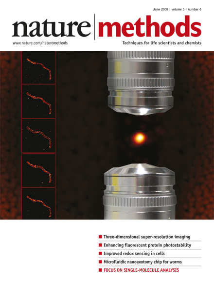
Artist's rendition of isoSTED microscopy based on an idea by Roman Schmidt, Alexander Egner and Stefan Hell. The opposing objectives and resulting spherical imaging spot used in isoSTED microscopy are shown beside a vertical stack of isoSTED images of Tom20 protein in a mitochondrion after immunofluorescence labeling. Cover by Erin Boyle. Article, p539, News and Views p471

Editorial

  • Biologists are increasingly interested in single-molecule approaches. In this issue, a Focus provides a biologist's guide to this relatively new field, and two papers present advances in nanoscale visualization.

    Editorial

    Advertisement

Top of page ⤴

Correspondence

Top of page ⤴

Research Highlights

  • Researchers generated a high-resolution snapshot of the epigenome of Arabidopsis thaliana by constructing and integrating the methylome, transcriptome and small RNAome using next-generation sequencing.

    • Michelle Pflumm
    Research Highlights
  • A 3D laser scanning microscopy method requiring no moving parts promises to expand the in vivo study of fast neuronal signaling at cellular and subcellular levels.

    • Daniel Evanko
    Research Highlights
  • By adding new connections between unrelated genes to a gene network, researchers can investigate network robustness and evolvability.

    • Allison Doerr
    Research Highlights
  • Virtual composite images generate a map of gene expression in Drosophila embryos.

    • Natalie de Souza
    Research Highlights
  • Synthetic peptides that can enter the cell and localize to mitochondria are promising tools for delivering cargo to the organelle.

    • Irene Kaganman
    Research Highlights
Top of page ⤴

News & Views

Top of page ⤴

Review Article

Top of page ⤴

Perspective

Top of page ⤴

Brief Communication

  • The ability to image thick volumes with invariant high axial and lateral resolution is a challenge for existing super-resolution fluorescence microscopy techniques. The combination of a double-plane detection scheme with fluorescence photoactivation microscopy (FPALM) allows three-dimensional sub-diffraction resolution imaging of samples as thick as whole cells.

    • Manuel F Juette
    • Travis J Gould
    • Joerg Bewersdorf
    Brief Communication
  • Caenorhabditis elegans is an ideal model organism for studying nerve regrowth and functional recovery after in vivo axotomy, but its high mobility makes such experiments challenging. A microfluidic device capable of transient immobilization of individual worms for high-resolution imaging and laser-based nanoaxotomy is described.

    • Samuel X Guo
    • Frederic Bourgeois
    • Adela Ben-Yakar
    Brief Communication
  • To date, the only way to array proteins with high density and high content has been to print purified proteins on a microarray surface. The next generation of nucleic acid programmable protein arrays (NAPPA) now allows thousands of proteins to be produced in situ on a microarray.

    • Niroshan Ramachandran
    • Jacob V Raphael
    • Joshua LaBaer
    Brief Communication
Top of page ⤴

Article

  • A fluorescence microscope relying entirely on focused light allows the generation of spherical focal fluorescence spots much smaller than the wavelength of light. This development, termed isoSTED, overcomes the resolution limitation imposed by the diffraction of light and permits three-dimensional nanoscale imaging inside cells with common fluorophores.

    • Roman Schmidt
    • Christian A Wurm
    • Stefan W Hell
    Article
  • Improved photostability of fluorescent proteins would benefit many applications but is usually an afterthought in selection screens. Setting photostability as the primary selection criterion in screens for improved fluorescent proteins yielded highly photostable variants of existing orange and red fluorescent proteins without compromising other beneficial characteristics.

    • Nathan C Shaner
    • Michael Z Lin
    • Roger Y Tsien
    Article
  • Analysis of intracellular redox-based processes is constrained by the limited choice of appropriate biosensors. Fusion of human glutaredoxin-1 to an existing redox-sensitive GFP results in a ratiometric biosensor that allows rapid and sensitive dynamic imaging of glutathione redox potential in living cells.

    • Marcus Gutscher
    • Anne-Laure Pauleau
    • Tobias P Dick
    Article
  • Many extracellular receptors are organized into complexes that may have functional implications. A combination of snap-tag protein labeling technology with time-resolved fluorescence resonance energy transfer (FRET) provides a method for the systematic analysis of higher-order protein-protein interactions on the surface of living cells.

    • Damien Maurel
    • Laëtitia Comps-Agrar
    • Jean-Philippe Pin
    Article
Top of page ⤴

Technology Feature

  • In less than five years the Nano/Bio Interface Center at the University of Pennsylvania has gone from an idea to a nationally funded nanotechnology center. A look inside reveals how they have taken a collaborative approach to technology development.

    • Nathan Blow
    Technology Feature
Top of page ⤴

Erratum

Top of page ⤴

Advertising Feature: Application Note

Top of page ⤴

Focus

  • Specially commissioned Reviews and a Perspective provide a guide to the maturing field of single-molecule analysis and two original research papers describe complementary advances in nanoscale visualization.

    Focus
Top of page ⤴

Search

Quick links