Thank you for visiting nature.com. You are using a browser version with limited support for CSS. To obtain
the best experience, we recommend you use a more up to date browser (or turn off compatibility mode in
Internet Explorer). In the meantime, to ensure continued support, we are displaying the site without styles
and JavaScript.
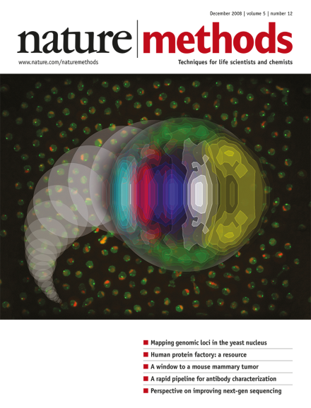
A probabilistic map of the locations of several genomic loci within the yeast nucleus, overlaid onto fluorescence images of yeast, from which such maps can be derived. Cover design by Erin Boyle, based on images provided by Christophe Zimmer. Aricle p1031
Sequencing technology is now advanced enough to decode individual human genomes. Will it prove to be better than existing methods for discovering the genetic basis of human phenotypic variation?
Researchers describe a method for protein identification and quantification based on electron-vibration-vibration two-dimensional infrared spectroscopy.
To scale up the production and complexity of DNA nanostructures, researchers enlist the help of Escherichia coli to replicate and assemble them in vivo.
An algorithm for identifying allosteric mechanisms allows researchers to assemble a functional multidomain protein and may offer new evolutionary insights.
Comprehensive sets of clones and improved high-throughput methods for production of functional proteins now allow proteome-scale in vitro experiments on nearly 15,000 human genes.
Efficient methods to characterize the binding properties of affinity reagents are required. A combination of bacterial surface display, flow cytometry and pyrosequencing is now used for high-speed mapping of the epitopes recognized by antibodies.
A collection of 33,275 human Gateway entry clones and complementary in vitro protein expression methodologies are described that allow proteome-scale production of human proteins. This 'human protein factory' was validated by expression of 13,364 human proteins and assessment of activity in a variety of assays.
The combination of a glass window placed on top of a mouse mammary gland with photoswitchable fluorescent protein labeling of implanted tumor cells allows tumor-cell tracking over multiple imaging sessions in orthotopic tumors. Results show the existence of two distinct microenvironments with different tumor-cell invasion and intravasation characteristics.
The algorithm Sylamer finds over- or underrepresented nucleotide motifs, such as microRNA seeds, in a gene list ranked according to expression levels and thus establishes whether a microRNA is directly affecting gene expression.
A simple modification to the optical configuration used for fluorescence photoactivation localization microscopy (FPALM) allows the fluorescence anisotropies of each individual molecule in a nanoscale image to be measured. The method was used to obtain position and orientation information for fluorescently labeled actin or hemagglutinin molecules in fixed fibroblasts.
The spatial organization of the genome within the eukaryotic cell nucleus is not random. Automated imaging of thousands of live yeast is now used to build high-resolution probabilistic maps of the locations occupied by individual loci.
An efficient pipeline for mapping antibody epitopes is presented. Combining bacterial surface display of peptide libraries, flow cytometric sorting, and pyrosequencing, the approach is amenable to a high-throughput format and should find future application in whole-proteome studies.
Extension of multicolor three-dimensional stochastic optical reconstruction microscopy (STORM) allows super-resolution fluorescence imaging of whole cells and quantitative characterization of subcellular structures and their spatial relationships. This was demonstrated by imaging the entire mitochondrial and tubulin networks in cells.
Co-patterning of a membrane protein bait and a fluorescently labeled prey is used to examine protein-protein interactions in a semiautomated fashion in living cells. Photobleaching experiments and single-molecule imaging further allow dynamic studies of the interaction.
With increasing numbers of well-characterized stem cell lines and improved culture and differentiation technologies, more scientists are testing the waters of stem cell research.