Thank you for visiting nature.com. You are using a browser version with limited support for CSS. To obtain
the best experience, we recommend you use a more up to date browser (or turn off compatibility mode in
Internet Explorer). In the meantime, to ensure continued support, we are displaying the site without styles
and JavaScript.
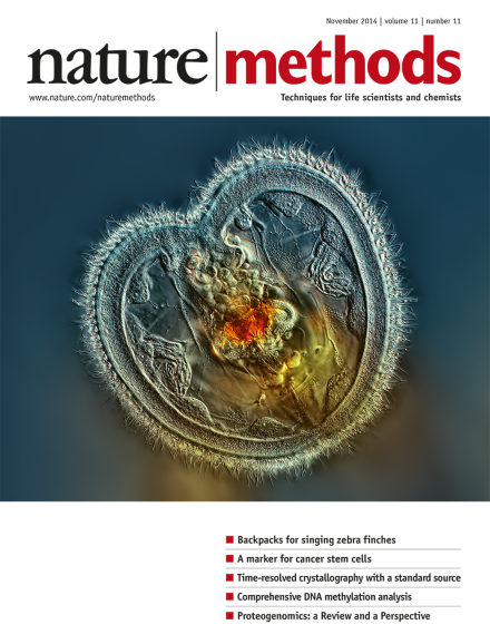
Image of a rotifer showing the mouth interior and heart-shaped corona, photographed using differential interference contrast microscopy (40× magnification) by Rogelio Moreno, Panama City, Panama. Winner of the 2014 Nikon Small World photomicrography contest (reprinted with permission from Nikon).
Imaging of electrical activity in vivo in Caenorhabditis elegans is possible with the improved genetically encoded voltage sensors Archer1 and Archer2.
Researchers grow genetically stable organoids from mouse and human prostate cells in the lab and establish patient-derived lines that model prostate cancer.
Electrodes and electrode arrays to record from neurons come in an increasing number of shapes and sizes, and engineers are continuously adding capabilities.
Tumor-specific peptides missed in standard mass spectrometry–based workflows can be identified by integrating genomic information to interpret proteomic data. As discussed in this Perspective, an onco-proteogenomic approach to study tumor biology could have a substantial impact on cancer research. Also in this issue, Nesvizhskii reviews proteogenomic technology.
A proteogenomic approach to analyzing mass spectrometry–based proteomic data enables the discovery of novel peptides, provides peptide-level evidence of gene expression, and assists in refining gene models. Strategies for building custom sequence databases, applications benefitting from a proteogenomic approach, and challenges in interpreting data are discussed in this Review. Also in this issue, Alfaro et al. discuss the use of proteogenomic approaches for studying cancer biology.
A microfluidic chip is used to construct a microarray of proteins, each labeled with a dockerin tag, for high-throughput single-molecule force spectroscopy experiments using a single cohesin-modified cantilever.
A method based on the Hadamard transform is shown to enable time-resolved X-ray crystallography measurements of protein dynamics at standard synchrotron sources.
Communications between animals such as zebra finches can be discriminated with back-attached acceleration recorders. In contrast to microphones, these devices record the carrier's signals only, allowing a more precise analysis of individual vocalizations during social interactions.
The CONCOCT software performs unsupervised binning of metagenomic contigs across multiple samples to allow better genome reconstruction from microbial communities.
A model that incorporates the quantitative relationship between microRNA and the expression of its target gene achieves predictable and robust genetic circuits.
Designer ribozymes show protein-responsive translational control and can be combined with transcriptional control elements to program complex gene circuits.
This paper reports an autofluorescent signal in cancer stem cells within epithelial tumors and describes its use as a marker to isolate and study these cells.
This paper reports a combination of two small molecules for very efficient mouse cell reprogramming to induced pluripotency, achieving close to 100% reprogramming within a few days for some cell types.
Quantum dots sequentially loaded into cells are used to generate barcodes that can identify thousands of individual cells within a population and that can be used to track cells over many hours.