Volume 23
-
No. 12 December 2017
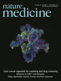
Broutier et al. generated human primary liver cancer (PLC)-derived organoids from patient tissues. These PLC-derived organoids recapitulate all the features of human PLCs, from histological architecture to genetic and transcriptomic traits, and are amenable as a platform for drug screening. The cover image depicts a confocal projection of hepatocellular carcinoma (HCC)-derived organoids, with the HCC marker AFP in white and Hoechst in blue. Image credit, Laura Broutier.
-
No. 11 November 2017
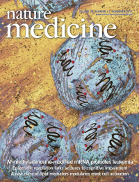
N6-methyladenosine (m6A) methylation is the most abundant modification in mRNA. Vu et al. (p 1369) find that the m6A-forming enzyme METTL3 is upregulated in acute myeloid leukemia compared to normal immature blood cells and that METTL3 promotes leukemia by controlling the stability and translation efficiency of its target mRNAs. The cover image depicts myeloid leukemia cells containing mRNAs with the m6A modification (red). Image credit: Ly P. Vu and artwork by Lewis Long.
-
No. 10 October 2017
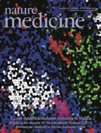
Understanding immunity to malaria is crucial to the development of effective vaccines and immune-modulating therapies. In this issue (p 1220), John Harty and colleagues report that regulatory T cells inhibit conventional T cell and germinal center B cell responses through the inhibitory receptor CTLA-4, affecting antimalarial immunity. The cover image depicts a germinal-center reaction in the spleen of a mouse infected with Plasmodium yoelii, after treatment with anti-CTLA-4. CD4+ T helper cells (green), B cells (blue), germinal-center B cells (white) and plasmablasts (red) are shown. Image credit: Samarchith P. Kurup and the University of Iowa Central Microscopy Research Facility. Artwork by Erin Dewalt.
-
No. 9 September 2017
Major depressive disorder (MDD) is a debilitating syndrome that affects men and women differently. Labonté and colleagues (p 1102) show that males and females with MDD exhibit widely different transcriptional signatures across brain regions. By translating these findings into an animal model of stress, the team modulated the activity of gene networks, demonstrating their contributions to neuronal activity and stress susceptibility in a sex-specific fashion. Overall, the results suggest that MDD in males and females may arise from sex-specific transcriptional alterations, which ultimately converge by driving similar symptomatic features. Illustration credit: Frederic Cantin, Labonté Lab.
-
No. 8 August 2017
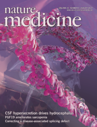
In a rat model of intraventricular hemorrhage, Karimy et al. (p 997) investigate the role of TLR4–NF-κB-dependent inflammation in the development of post-hemorrhagic hydrocephalus. Inflammation at the choroid plexus epithelium (CPE) induces hypersecretion of cerebrospinal fluid (CSF) through phosphorylation of the SPAK–NKCC1 ion co-transporter complex. Therapeutics that attenuate this inflammatory response restore basal rates of CSF secretion and subsequent pathologic enlargement of the cerebral lateral ventricles. The cover image depicts the CPE (red) secreting CSF (gray spheres) within the cerebral ventricles. Image credit: Ella Maru Studio, Alexander Tokarev, and the Kahle laboratory.
-
No. 7 July 2017
By performing stepwise treatment with growth factors and WNT pathway modulators, Crespo et al. (p 878) design a protocol to generate colonic organoids from human induced pluripotent stem cells (iPSCs). They apply this approach to derive organoids from iPSCs from patients with familial adenomatous polyposis (FAP). A drug screen using these organoids indicates that the antibiotic geneticin suppresses abnormal WNT signaling in organoids derived from FAP cells bearing ATM mutations. The cover image depicts a confocal projection of FAP-iPSC-derived colonic organoids, with colon marker CDX2 and proliferation marker CD1 false-colored in green and red, respectively. DAPI, blue. Image credit, Miguel Crespo. Artwork, Erin Dewalt.
-
No. 6 June 2017
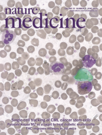
Using a new method enabling simultaneous whole transcriptome sequencing and detection of the BCR-ABL fusion gene in single cells, Giustacchini et al. (p 692) track the evolution of cancer stem cells in patients with chronic myeloid leukemia. In addition to characterizing rare persistent tumor cells present in patients on prolonged therapy, this approach revealed disease-associated alterations to non-malignant hematopoietic cells. The cover image depicts a blood smear from a patient with CML, with a single CML cell false-colored. Image credit, Tiago C Luis, Nikolaos Sousos, Connor Sweeney and Alice Giustacchini. Artwork, Erin Dewalt.
-
No. 5 May 2017
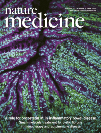
West et al. (p 579), find that the cytokine oncostatin M (OSM) and its receptor are upregulated in the inflamed intestinal tissue from patients with inflammatory bowel disease (IBD). High OSM expression was associated with resistance to tumor necrosis factor (TNF)-neutralizing therapy. OSM blockade or genetic deletion attenuated disease severity in a mouse model of anti-TNF resistant IBD, suggesting that OSM may be a promising therapeutic target. Image depicts inflamed mouse colon mucosa, with an expanded population of intestinal stromal cells identified by expression of podoplanin (green). Epithelium is identified by expression of EpCAM (magenta); cyan, nuclei. Image credit, Samuel Bullers.
-
No. 4 April 2017
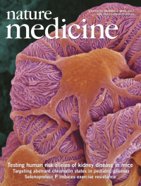
In this issue (p 429), Katalin Susztak and colleagues tested whether two previously identified human risk alleles of APOL1 cause kidney disease when expressed as transgenes in mice. They find that expression of either risk allele, unlike that of a reference nonrisk allele, specifically in podocytes is sufficient to cause such disease in mice. Neither risk allele caused pathology when expressed in renal tubules. The cover shows a pseudocolored scanning electron micrograph of a glomerulus. Image credit: Dennis Kunkel Microscopy/Science Source.
-
No. 3 March 2017
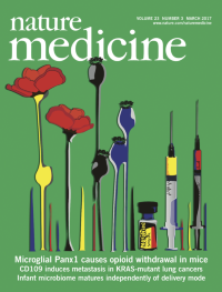
The opium poppy is a natural source of opioids such as morphine. Although they are revered for their potent pain-relieving effects, when used chronically, opioids can lead to analgesic tolerance, hyperalgesia and withdrawal. Burma et al. (p 355) show that pannexin-1 (Panx1) channels on spinal microglia are essential for opioid withdrawal, and that blocking Panx1 can alleviate opioid withdrawal in rodents. Puig and Gutstein (p 272) discuss these findings alongside those recently reported by Corder et al. in the February issue of Nature Medicine. Illustration credit: Nicole Burma, Trang laboratory.
-
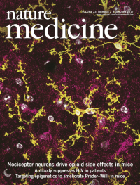
No. 2 February 2017
Corder et al. establish that μ opioid receptors (MORs) on peripheral nociceptor neurons, but not microglia, are responsible for opioid-related side effects of tolerance and opioid-induced hyperalgesia. They further demonstrate that a commercially available, peripherally restricted opioid antagonist, methylnaltrexone bromide, mitigates these side effects when co-administered with morphine. Image depicts mouse spinal cord with Oprm1 mRNA (coding for MOR, fuchsia) concentrated in neurons but not microglia (CD11b, yellow). Image credit: Vivianne Tawfik, Scherrer laboratory.
-
No. 1 January 2017
Workman et al. describe a method to generate intestinal organoids bearing a functional enteric nervous system (ENS), from human pluripotent stem cells. They apply this method to reveal insight into the molecular alterations associated with Hirschsprungs disease. Image depicts an organoid with an ENS. Red, intestinal epithelium; green/yellow, ENS neurons. Image credit, Maxime Mahe.