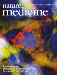
Volume 21
-
No. 12 December 2015
In this issue, we present a series of Reviews and Perspectives on aging. Loss of proteostasis is a hallmark of aging and may be activated by treatment with rapamycin which results in autophagy as observed in cultured neuronal cells. Autolysosomes are shown in red, and nuclei in blue. Image credit: Antonio Diaz
Focus
-
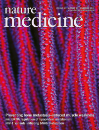
No. 11 November 2015
Waning and Mohammad et al. (p 1262) show that elevated levels of transforming growth factor-β (TGF-β), released as a consequence of tumor induced osteolysis, result in muscle weakness, even before the loss of skeletal muscle mass. Blocking TGF-β or its downstream mediators prevents this pathology in mouse models. The cover image depicts red whip coral (Ellisella ceratophyta), reminiscent of skeletal muscle fibers. Image credit: Theresa Guise.
-
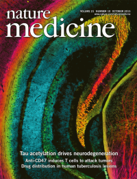
No. 10 October 2015
In this issue, Min et al. (p 1154) identify acetylation of tau at site K174 as an early pathological change in human Alzheimer's Disease (AD) brain tissue and show that this acetylation exacerbates tau-mediated neurodegeneration and memory impairments in mice. Furthermore, pharmacological inhibition of tau acetylation can ameliorate these phenotypes in a mouse model of AD/FTD (frontotemporal dementia). The cover image depicts immunostaining for AC312-positive acetylated tau (red and magenta) and MC1-positive pathological tau (blue and green) in the hippocampus of the PS19 transgenic mouse model of AD/FTD. Image credit: the original image is by Chao Wang, and the artwork is by Connor Ludwig.
-
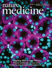
No. 9 September 2015
In this issue, Lovisa et al. (p 998) and Grande et al. (p 989) show that during renal fibrosis a partial epithelial-to-mesenchymal program results in loss of normal renal parenchyma function, contributing to the fibrotic process; this program can be genetically targeted to reverse established disease. The cover image depicts immunostaining of the organic anion transporter OAT3/SLC22A6 (pink) and nuclei (DAPI, cyan) in normal murine kidney tissue. Image credit: Sara Lovisa and Valerie LeBleu.
-
No. 8 August 2015
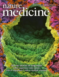
Two papers in this issue reveal new insight into lung regeneration. Rosen et al. (p 869) report that preconditioning by sublethal radiation allows better engraftment of embryonic lung tissue into the injured lung of adult mice, leading to improved lung repair. Liu et al. (p 866) find by lineage tracing that c-kit+ cells do not, as previously reported, contribute to lung epithelium during homeostasis or repair after injury, but rather maintain an endothelial fate. Image shows a colored scanning electron micrograph of normal lung tissue, including the lumen of a blood vessel (yellow) lined with endothelial cells, as well as alveoli air sacs lined with capillaries (blue and pink). Image credit: Professor Pietro M. Motta / Science Source.
-
No. 7 July 2015
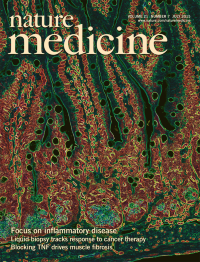
In this issue, we are proud to feature a collection of articles on inflammatory disease (pp 669740). The cover image depicts the epithelium that lines the mouse gastrointestinal tract and influences immunity. The cell membrane of small intestinal epithelial cells is visualized in green with the nuclei shown in orange. Paneth cell granules at the base of the crypts are marked in blue. Image courtesy of Mario Noti, Gregory F. Sonnenberg and David Artis. Artwork by Erin Dewalt.
Focus
-
No. 6 June 2015
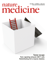
Two papers in this issue widen the spectrum of known mechanisms used by tumors to escape therapy. By sequencing cell-free plasma DNA from lung cancer patients treated with an epidermal growth factor receptor (EGFR) inhibitor, Thress et al. reveal a new EGFR-resistance mutation. Whole-exome sequencing of samples from patients with acute lymphoblastic leukemia led Li et al. to mutations in the phosphoribosyl pyrophosphate synthetase 1 gene (PRPS1) and a new chemotherapy-resistance mechanism. Cover image: stuartmiles99/iStock/ Thinkstock
-
No. 5 May 2015
Using ClonTracer, a high-complexity genetic barcoding system with the capacity to uniquely label millions of cells with different DNA barcode sequences, Bhang et al. (p 440) show that in preclinical cancer models, the majority of resistant clones already preexist as rare subpopulations prior to therapy and selectively escape therapy. This technology can be used to prioritize combination therapies with distinct resistance mechanisms with the goal of preventing the emergence of resistance. Artwork by Alan Abrams.
-
No. 4 April 2015
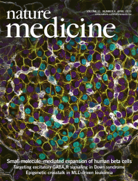
Using a high throughput screen, Wang et al. (p 383) designate the small molecule harmine a human beta cell mitogen. Acting through the dual-specificity tyrosine-regulated kinase-1a (DYRK1A), harmine boosts beta cell proliferation and improves glycemic control in several preclinical in vivo models. Image depicts immunostaining of beta cells in the adult mouse islet of Langerhans. Image credit: Erik Bader and Heiko Lickert. Artwork by Erin DeWalt.
-
No. 3 March 2015
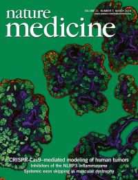
Using CRISPR-Cas9–based genome editing technology, Sato and colleagues (p 256) introduce various combinations of mutations associated with human intestinal tumors into organoids derived from healthy human intestinal epithelium. This approach— which might be applied more broadly with other mutations—reveals insight into the number and types of mutations needed for tumorigenesis and acquisition of an invasive phenotype. Image depicts engineered organoids; apical membrane (blue), basal membrane (red) and nucleus (green). Image credit: Yuki Ohta and Toshiro Sato. (In the version of the cover caption initially published, the definitions of the green and blue staining were reversed. Green represents nuclear staining, and blue represents the apical membrane. The error has been corrected in the HTML version of the caption as of 25 March 2015.)
-
No. 2 February 2015
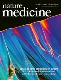
Hundreds of susceptibility loci have previously been associated with autism spectrum disorder (ASD) suggesting the disease is genetically heterogeneous. In this issue, Stephen Scherer and colleagues report a whole-genome sequencing study of 85 quartet families (parents and two ASD-affected siblings). The cover image, courtesy of Linden Gledhill and Autism Speaks, depicts the growing face of a liquid crystal formed from an evaporating aqueous solution of DNA (specifically, the 14 base pair double stranded DNA palindrome fragment 5-ACGCGAATTCGCGT-3). The blue color is due to birefringence imaged using cross-polarized light microscopy (magnification, x100).
-
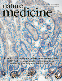
No. 1 January 2015
Davis et al. (p 62) report that disruption of key homeostatic morphogen signaling gradients in the intestine results in the expansion of a population of proliferating progenitor cells outside of the crypt base stem cell niche. These cells do not express the intestinal stem cell marker Lgr5, yet form ectopic crypts on the villus, acquire somatic mutations and initiate tumorigenesis. The image shows immunohistochemical staining for an Lgr5-EGFP reporter gene in the small intestine of a mouse expressing a transgene encoding the bone morphogenetic protein antagonist Grem1 in intestinal epithelial cells. Lgr5-EGFP positive cells (brown) are found at the base of the intestinal crypts, but not in ectopic crypt foci on the villus. Original image by Hayley Davis.