
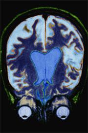
 Spotting Alzheimer’s in the brain before symptoms start would allow treatment to begin years earlier than is currently possible.© Punchstock
A brain scan developed in mice could herald a safe and affordable way to screen patients for Alzheimer's disease before they show any symptoms.
Currently, doctors can diagnose the disease only after patients develop traits such as forgetfulness and confusion. But 10 to 20 years before these symptoms appear, toxic clumps of protein called amyloid plaques form in the brain.
Researchers have characterized these tiny plaques in brain tissue after death, but they have struggled to capture images of them in living patients.
Now Takaomi Saido and colleagues at the RIKEN Brain Science Institute in Wako City, Japan, have developed a way to view these plaques in the brains of live mice using MRI, a magnetic imaging technique widely available in hospitals.
High contrast
Previous MRI studies used signals from hydrogen, which occurs naturally in the body. But this made it hard to see the pinpoint-sized plaques against the background noise of other structures in the brain.
To turn up the contrast, Saido and his colleagues adapted a non-toxic compound that is known to bind to the plaques. They substituted one atom of the compound with a derivative of fluorine that is not naturally found in mice or humans. It produces a distinctive magnetic signal that is easy to detect using MRI.
When the researchers injected the compound into mice that had a condition equivalent to Alzheimer's, it bound successfully to the plaques in their brains.
"We were able to get images with very high contrast," says Makoto Higuchi, one of the authors of the study. The team has published the result in Nature Neuroscience1.
"There's a significant gain," agrees Scott Small, a neurologist at the Columbia University College of Physicians and Surgeons in New York. "There's essentially no noise."
Into the clinic
Imaging amyloid plaques in patients would offer doctors a chance to diagnose and treat Alzheimer's disease years earlier than is possible at the moment.
A radioactive imaging technique called PET scanning is being tested in clinical studies, but its resolution is limited to millimetres, and individual plaques are at least ten times smaller than that.
ADVERTISEMENT
The use of MRI could be a better option, researchers say. In addition to its high resolution, it is cheaper, more widely available and safer than PET, which requires the patient to be injected with a radioactive substance.
Having shown that the MRI method works in mice, the researchers hope to be able to test the technique in humans within a few years. "Our technique is not immediately applicable, but it certainly shows the way," Higuchi says.
Before moving to clinical trials the team will need to refine the technology and the design of the compound to be absolutely sure of the method's safety. "It's a nontrivial hurdle, but it's surmountable," says Small.
