Thank you for visiting nature.com. You are using a browser version with limited support for CSS. To obtain
the best experience, we recommend you use a more up to date browser (or turn off compatibility mode in
Internet Explorer). In the meantime, to ensure continued support, we are displaying the site without styles
and JavaScript.
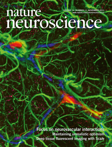
Interactions between neurons, glia and vasculature are critical for the maintenance of normal brain function. We present a special focus on the growing field of neurovascular interactions comprising reviews and perspective articles highlighting the latest advances in our understanding of these interactions, with particular emphasis on their roles in health and disease. The cover depicts a confocal image composite of perivascular migration of implanted human glial progenitor cells, xenografted into the rodent brain, courtesy of Takahiro Takano, Maiken Nedergaard and Steven Goldman.135313631405
Blood vessels in the nervous system are not simply inert bystanders that only support the metabolic needs of neurons. We present a focus on neurovascular interactions that highlights our emerging knowledge of how these interactions shape neuronal function both in health and disease.
People tend to remain overly optimistic even when faced with information about a gloomy future. A study now shows that people are selectively worse at incorporating information about a worse-than-expected future. It also describes the learning signals in the brain that correlate with this bias.
Neurons form synapses with oligodendrocyte precursor cells (OPCs) that may control their maturation and myelination. Key signaling molecules regulating glutamate receptors at neuronal synapses also act in OPCs, but to opposite effect.
Patchy variation in odor-evoked electrical activity in the human olfactory epithelium is found to correlate with stimulus pleasantness. This finding depends on a new technique for recording directly from awake humans.
There remains an urgent need to develop new strategies and therapies to help protect the brain from ischemic cell death. In this perspective, the authors suggest that learning more about the mechanisms that underlie brain self-preservation and developing multifaceted approaches that act on multiple pathways involved in both cell death and neuroprotection may advance our efforts to treat stroke.
This perspective discusses newly discovered mechanisms leading to cellular ionic imbalances, as well as underappreciated signaling cascades that mediate cell death and that may add to the traditional glutamatergic mechanisms to which ischemic brain injury is ascribed. An integrated consideration of such new mechanisms may aid in formulating better therapies.
Brain tumor stem cells (BTSCs) stimulate angiogenesis and may also directly contribute to tumor vasculature. The authors review the codependence of BTSCs and the perivascular niche and how this may inform new therapeutic approaches.
Blood vessels in the CNS have traditionally been considered neutral bystanders that passively adapt in response to the needs of neural cells. This review surveys recent evidence that blood vessels actively participate in the pathogenesis of neurological disorders and the implications of this work for therapy.
Maturation of adult-born neurons in the dentate gyrus is known to require GABAergic input. Here the authors show that a subtype of interneurons, namely neurogliaform cells, acts as the primary source of GABA for newborn neurons in mouse dentate gyrus.
The authors generated knock-in mice of the AMPAR auxiliary subunit TARP that lack the C-terminal PDZ ligand. They found that synaptic transmission and AMPAR were reduced without changes in extrasynaptic AMPAR expression, but LTP was unaltered.
Using hemisphere-specific optogenetic activation of hippocampal fibers, this study finds that the magnitude of long-term potentiation in CA1 neurons depends on whether afferents originate in left or right CA3.
This study shows that lesioning a rat's amygdala affects only familiarity-based recognition, having no effect on recollection-based recognition, and further dissociates the role of medial temporal lobe structures mediating recognition memory.
Using two-photon microscopy in mice, the authors find that the number of cortical spines increases in adolescent mice while they are awake and decreases while they are asleep.
Examining the role of ephrin-B3 in dendritic and synaptic development in vivo, this study finds that ephrin-B3 functions postsynaptically as a receptor to initiate reverse signaling events that organize dendritic branching complexity, spine maturation and formation of functional synapses.
The authors studied the dynamic regulation of Ca2+-permeable AMPARs (CP-AMPARs) in oligodendrocyte precursor cells. Group 1 mGluRs regulate CP-AMPARs via a pathway that requires intracellular Ca2+, PI3 kinase, PICK-1 and JNK. Purinergic receptor activation decreases CP-AMPAR expression.
Cocaine can easily cross the placental and fetal blood-brain barrier, and in utero exposure to cocaine can cause lasting behavioral changes in postnatal periods. Here, Bellone et al. studied the physiological and circuit level mechanism behind the consequence of in utero cocaine exposure and found a postnatal synaptic maturation defect of excitatory input to the dopaminergic neurons in the ventral tegmental area of mice. In particular, they found that late embryonic in utero cocaine exposure causes a delay in AMPAR/NMDAR switch in early postnatal mouse brain.
This study examines the contribution of a specific phosphatidylinositol 3-kinase (PI3K) isoform, namely PI3Kγ, to hippocampal synaptic plasticity and behavior. The authors find that the loss of PI3Kγ can specifically impair NMDA receptor–mediated long-term depression and cognitive functions that rely on behavioral flexibility.
Inserting a recording electrode into the nostrils of human volunteers allowed the authors to record neural activity directly from the olfactory epithelium, and measures of olfactory perception, all from the same individuals. This uncovered a non-uniform patchy organization of the receptive surface, which was organized in part according to the perception of odorant pleasantness.
One mechanism by which medial prefontal cortex (mPFC) exerts cognitive control is thought to involve the subthalamic nucleus (STN), which acts as a temporary brake on behavior. Here the authors found increases in mPFC and STN theta power as a function of decision conflict. Increases in mPFC theta power predicted increased decision thresholds. STN deep brain stimulation reversed this relationship, resulting in impulsive choice.
The authors examine the neural circuitry causally involved in normative, fairness-related decisions by generating a temporarily diminished capacity for costly normative behavior through non-invasive brain stimulation. Their findings suggest that a prefrontal network, the activation of rDLPFC and pVMPFC and the connectivity between them, facilitates costly normative decisions.
This study reports that people are worse at incorporating negative information when updating their beliefs. Correspondingly, neural activity encodes desirable information updates, but there is weaker encoding of unexpectedly undesirable information.
The authors describe a chemical approach for imaging deep into fixed brain tissue using Scale, a solution that renders biological samples transparent, but preserves fluorescent signals. This technique allows for imaging at unprecedented depth and at subcellular resolution, and makes three-dimensional reconstruction of neural networks possible without serial sectioning.
Blood vessels in the nervous system are not simply neutral bystanders that only support the changing needs of neurons. Nature Neuroscience presents a focus on neurovascular interactions that highlight our emerging knowledge of how these interactions shape neuronal function both in health and disease. The focus includes reviews and perspectives written by experts in the field discussing how blood vessels and angiogenic molecules actively participate in the pathogenesis of various neurological disorders. Reviews also critically evaluate our advances in understanding how blood vessels interact with neural stem and progenitor cells, brain tumor interactions with the neurovasculature, and the consequences for disease when these interactions are affected.