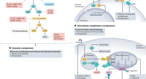
Cardiac Fabry's disease: an unusual cause of left ventricular hypertrophy
José Pedro L Nunes*, Ovídio Costa, Maria do Sameiro Faria, Pedro Bernardo Almeida and Lúcia Lacerda About the authors
Correspondence *Faculdade de Medicina do Porto, Alameda Prof Hernâni Monteiro, 4200 Porto, Portugal
Email jplnunes@med.up.pt
Vanderbilt University School of Medicine is pleased to provide the opportunity to earn one Category 1 credit toward the American Medical Association's Physician's Recognition Award (AMA PRA). Please refer to the AMA website awards page for further information on this award.
To obtain credit, please go to the Vanderbilt CME website and answer five multiple-choice questions. Only one answer is correct for each question. You must score 80% or higher to obtain credit. Questions for each quiz are available for 12 months.
To complete the CME questions you must have an account in the Vanderbilt CME database. If you do not have a Vanderbilt CME ID you will be asked to register and provide personal data that will be used to (1) enter the credit you earned in the Vanderbilt CME database and (2) print a certificate for your files that you can use to provide evidence that you earned CME credit. (Note: Your personal data will be used only for this purpose. If you have questions or need more information, send an email to cme@vanderbilt.edu).
Summary
Background A 64-year-old male was observed as an outpatient with atypical, non-exercise-induced chest pain and palpitations. He had arterial hypertension and marked concentric left ventricular hypertrophy. After 2.5 years of antihypertensive drug therapy the patient's blood pressure had returned to normal, but his left ventricular hypertrophy was unchanged.
Investigations Electrocardiography, transthoracic echocardiography, myocardial perfusion scintigraphic imaging, measurement of
-galactosidase A activity, gene sequencing, brain MRI, carotid artery ultrasonography, biochemical renal evaluation and cardiac Doppler tissue imaging.
Diagnosis Cardiac Fabry's disease.
Management Losartan, hydrochlorothiazide, low-dose aspirin and bisoprolol. The patient is expected to begin enzyme replacement therapy.
The case
A 64-year-old man presented as an outpatient with atypical, non-exercise-induced chest pain and palpitations. He had no personal history of acroparesthesia, chronic pain, gastrointestinal problems, skin lesions, stroke or any other notable conditions. His mother had experienced sudden cardiac death at the age of 42 years, but his family history was otherwise unremarkable.
Electrocardiography of the patient showed sinus rhythm, normal PR interval (143 ms), left anterior fascicular block, high voltage of the QRS complex, and abnormal repolarization, including negative T-waves in the left precordial leads (Figure 1). Transthoracic echocardiography demonstrated marked concentric left ventricular hypertrophy and normal left ventricular systolic function. Myocardial perfusion scintigraphic imaging showed a normal perfusion pattern, and a left ventricular ejection fraction of 49%. Arterial hypertension was noted (170/100 mmHg), and the patient was started on losartan (50 mg/day), hydrochlorothiazide (25 mg/day) and low-dose aspirin (100 mg/day).
Figure 1 Electrocardiogram recorded at presentation, demonstrating left ventricular hypertrophy with strain pattern.
Owing to the high voltage of the QRS complex, electrocardiography was performed at the 5 mm/mV scale. The PR interval was 143 ms.
Full figure and legend (23K)Figures & Tables indexDownload Power Point slide (227K)At 1 year after presentation the patient's clinical status had completely stabilized and he was asymptomatic. Bisoprolol (5 mg/day) was added to the therapeutic regimen. A further 1.5 years later his blood pressure had returned to normal (120/70 mmHg), but echocardiography showed that his left ventricular hypertrophy had not resolved, prompting a search for an alternative cause. A total of six echocardiographic evaluations were carried out by different cardiologists over this 2.5 year period and in the following 2 years. Dilation of the left ventricular chambers was not observed at any point, but hypertrophy was consistently noted to persist (septal thickness measurements ranged from 16 mm to 20 mm and posterior wall thickness from 15 mm to 19 mm).
Upon biochemical analysis, low
-galactosidase A activity was seen in the patient's plasma, leukocytes and cultured skin fibroblasts (Table 1). Gene sequencing of cultured skin fibroblasts showed a Phe113Leu mutation in GLA, the gene encoding
-galactosidase A.1 The patient had no cutaneous lesions of interest. Further evaluation included brain MRI and carotid artery ultrasonography—both of which yielded normal results. Ultrasonography, performed as part of a renal evaluation, showed small lesions that were suggestive of calcifications. The plasma creatinine level was 107.0
mol/l (1.21 mg/dl; normal range 48.6–97.2
mol/l [0.55–1.10 mg/dl]), and 24 h creatinine clearance was 1.3 ml/s (76.7 ml/min; National Kidney Foundation chronic kidney disease stage 2). Non-nephrotic proteinuria (0.164 g/24 h; normal range 0.020–0.150 g/24 h) was also noted.
Table 1 Results of the patient's biochemical analysis.
Full tableFigures & Tables indexDownload Power Point slide (219K)
Enzyme replacement therapy (ERT) was considered an appropriate option by both medical staff and the patient, and an evaluation of the case by the relevant regulatory authority was started. The patient continued to take losartan, hydrochlorothiazide, low-dose aspirin and bisoprolol, as described above.
Approximately 4.5 years after initial presentation the patient remains asymptomatic. Authorization has been granted for ERT, which he is expected to start soon. The most recent noninvasive echocardiographic evaluation indicated a persistent increase in diastolic left ventricular wall measurements, along with a binary appearance of the left ventricular endocardial border (Figure 2), as has been previously described.2 The septal thickness was 18 mm, posterior wall thickness was 17 mm, left ventricular end-diastolic diameter was 53 mm, left ventricular end-systolic diameter was 37 mm and left ventricular ejection fraction was 57%. Doppler tissue imaging showed an E/E' ratio (early transmitral flow velocity to early diastolic mitral annular velocity) of 10.4, suggestive of diastolic dysfunction. Disease in the kidneys had improved—repeat evaluation showed a plasma creatinine level of 89.3
mol/l (1.01 mg/dl), 24 h creatinine clearance of 1.6 ml/s (96.7 ml/min; National Kidney Foundation chronic kidney disease stage 1) and 24 h proteinuria of 0.110 g/24 h.
Figure 2 Transthoracic echocardiographic images obtained 4 years and 4 months after the patient's initial presentation.
(A) Apical four-chamber view, and (B) parasternal short-axis view, demonstrating marked concentric left ventricular hypertrophy and the binary appearance of the left ventricular endocardial border (arrows), as described in cardiac Fabry's disease elsewhere.2 Abbreviations: LA, left atrium; LV, left ventricle; RA, right atrium; RV, right ventricle.
Full figure and legend (17K)Figures & Tables indexDownload Power Point slide (221K)Discussion of diagnosis
Fabry's disease (also known as Anderson–Fabry's disease) was originally described in 1898.3, 4 The condition is characterized by a deficiency in the enzyme
-galactosidase A, which leads to accumulation of globotriaosylceramide in the lysosomes of various cells.5 The clinical features of Fabry's disease include angiokeratoma, pain and acroparesthesias, gastrointestinal symptoms, hypohidrosis, corneal opacities, impaired renal function, stroke, and left ventricular hypertrophy.6 Manifestation of the disease is associated with a marked decrease in
-galactosidase A activity.5 The clinical presentation is variable and depends on the systems affected; stroke, end-stage renal failure and isolated left ventricular hypertrophy are all possible manifestations of the condition. Although the disease was previously considered to be relatively rare (around 1 in every 55,000 live births), its true prevalence is probably underestimated.6
Over 400 different mutations in GLA have been described in Fabry's disease, most of which are simple missense mutations.6 The condition is X-linked, making the disease more common among males, although females can also be affected.6
Atypical forms of Fabry's disease, predominantly affecting the heart, were reported almost a century after the original descriptions of the condition.7, 8, 9, 10 Several other cardiac variants have since been described, and various genetic mutations have been implicated (see Table 2 for a non-exhaustive description). All seven of the patients with Fabry's disease presented by Nakao et al., and five of the six presented by Sachdev et al., had symmetrical hypertrophy, which suggests that cardiac Fabry's disease could present primarily as a type of concentric left ventricular hypertrophy and, rather than being a very rare disorder, might simply go unrecognized in many cases.11, 12
Table 2 Some relevant published studies on the cardiac variant of Fabry's disease, presented in chronological order.
Full tableFigures & Tables indexDownload Power Point slide (262K)
The underlying pathophysiology of the cardiac variant of Fabry's disease is incompletely understood, but it is clear that different mutations in GLA are implicated. The accumulation of glycosphingolipids in the heart might not be sufficient by itself to account for the ventricular hypertrophy, which suggests that other mechanisms could be at play.6
Pieroni et al. described a binary appearance of the left ventricular endocardial border—an echocardiographic hallmark for cardiac Fabry's disease—that probably reflects the endomyocardial accumulation of glycosphingolipids.2 This finding was found to have a sensitivity of 94% and a specificity of 100% for the detection of Fabry's disease, and was also seen in the present case (Figure 2). The relevance of this hallmark as a diagnostic method, however, remains unconfirmed.
Endomyocardial biopsy can be helpful in the diagnosis of cardiomyopathies of uncertain etiology, especially when a treatable condition is suspected, and can assist in diagnosing Fabry's disease among patients who are heterozygous for GLA (females). It can, however, be difficult to justify this procedure in all patients with Fabry's disease, particularly in males, as endomyocardial biopsy has a risk of complications and a clear survival benefit associated with treatment has yet to be demonstrated. For these reasons, the procedure was not performed in the present case.
Arterial hypertension was initially suspected to be responsible for the marked left ventricular hypertrophy seen in the current patient, but after it had been adequately controlled for a prolonged period the cause of the hypertrophy remained unclear. Hypertrophic cardiomyopathy was also considered as an early possible diagnosis, but this option, along with other obvious causes of hypertrophy, was also ruled out. Biochemical and genetic screening for Fabry's disease were performed on the basis of observation of concentric (non-asymmetric) left ventricular hypertrophy and the absence of any other reasonable clinical explanation. These tests demonstrated low levels of
-galactosidase A in plasma, leukocytes and cultured skin fibroblasts, and the Phe113Leu (phenylalanine changed to leucine at position 113) missense mutation in GLA.1 The diagnosis was thereby established as a cardiac variant of Fabry's disease, taking into consideration the absence of systemic features of the condition. Minor renal involvement was present, but left ventricular hypertrophy was considered to be the major clinical finding. It is uncertain whether arterial hypertension might have had a role in the development of renal dysfunction, which, curiously, ameliorated over time.
Treatment and management
ERT using recombinant
-galactosidase A is currently available and constitutes the core of medical therapy for Fabry's disease. This treatment is generally considered to be well tolerated—the major issue regarding its use lies in the uncertainty over the clinical benefit, particularly in atypical cases of Fabry's disease with a late onset. A randomized trial of ERT in a group of 82 patients (2:1 treatment:placebo ratio) showed promising results with the use of a composite end point of time to first clinical event (renal, cardiac, cerebrovascular or death).13 In another randomized trial, involving 15 adult male patients, left ventricular mass (as measured by MRI) was found to be reduced by ERT.14
There is a strong rationale in favor of ERT, despite the lack of conclusive evidence regarding its benefits (at least as far as survival is concerned). It must be recognized that the rarer the clinical or genotypic presentation of Fabry's disease, the less likely it will be that data from prospective randomized trials will be available in the foreseeable future. The discomfort caused by biweekly intravenous therapy must be taken into consideration; careful explanation to the patient of all relevant aspects of the treatment is, therefore, crucial. Ongoing research suggests that alternatives to ERT might be available in the future.15
In the present case, antihypertensive therapy led to complete normalization of blood pressure and resolution of symptoms, but left ventricular hypertrophy persisted. ERT was, therefore, considered a reasonable treatment option, and an application was made to the relevant regulatory authority. Authorization has recently been granted and therapy is expected to begin in the near future (agalsidase
, 1 mg/kg every 2 weeks). This is the first case of cardiac Fabry's disease for which ERT has been approved in Portugal.
Conclusions
A 64-year-old male was successfully treated for hypertension, but had left ventricular hypertrophy that persisted despite treatment. Biochemical and genetic tests confirmed a diagnosis of Fabry's disease—a rare condition characterized by a deficiency in the enzyme
-galactosidase A. Screening for this disease could be justified in cases of otherwise unexplained left ventricular hypertrophy, especially of the concentric type.
References
- Eng CM et al. (1997) Fabry disease: thirty-five mutations in the alpha-galactosidase A gene in patients with classic and variant phenotypes. Mol Med 3: 174–182 | PubMed | ISI | ChemPort |
- Pieroni M et al. (2006) Fabry's disease cardiomyopathy: echocardiographic detection of endomyocardial glycosphingolipid compartmentalization. J Am Coll Cardiol 47: 1663–1671 | Article | PubMed | ISI |
- Anderson W (1898) A case of angiokeratoma. Br J Dermatol 10: 113–117
- Fabry J (1898) Contribution to the knowledge of purpura haemorrhagica nodularis (purpura papulosa haemorrhagica hebrae) [German]. Arch Dermatol Res 43: 187–200
- Brady RO et al. (1967) Enzymatic defect in Fabry's disease: ceramidetrihexosidase deficiency. N Engl J Med 276: 1163–1167 | PubMed | ISI | ChemPort |
- Clarke JT (2007) Narrative review: Fabry disease. Ann Intern Med 146: 425–433 | PubMed | ISI |
- Tanaka H et al. (1988) Four cases of Fabry's disease mimicking hypertrophic cardiomyopathy [Japanese]. J Cardiol 18: 705–718 | PubMed | ChemPort |
- Elleder M et al. (1990) Cardiocyte storage and hypertrophy as a sole manifestation of Fabry's disease: report on a case simulating hypertrophic non-obstructive cardiomyopathy. Virchows Arch A Pathol Anat Histopathol 417: 449–455 | Article | PubMed | ISI | ChemPort |
- Nagao Y et al. (1991) Hypertrophic cardiomyopathy in late-onset variant of Fabry disease with high residual activity of alpha-galactosidase A. Clin Genet 39: 233–237 | PubMed | ISI | ChemPort |
- von Scheidt W et al. (1991) An atypical variant of Fabry's disease with manifestations confined to the myocardium. N Engl J Med 324: 395–399 | PubMed | ChemPort |
- Nakao S et al. (1995) An atypical variant of Fabry's disease in men with left ventricular hypertrophy. N Engl J Med 333: 288–293 | Article | PubMed | ISI | ChemPort |
- Sachdev B et al. (2002) Prevalence of Anderson–Fabry Disease in male patients with late onset hypertrophic cardiomyopathy. Circulation 105: 1407–1411 | Article | PubMed | ISI | ChemPort |
- Banikazemi M et al. (2007) Agalsidase-beta therapy for advanced Fabry disease: a randomized trial. Ann Intern Med 146: 77–86 | PubMed | ISI |
- Hughes DA et al. (2007) Effects of enzyme replacement therapy on the cardiomyopathy of Anderson-Fabry disease: a randomized, double-blind, placebo-controlled clinical trial of agalsidase-alfa. Heart [doi:doi: 10.1136/hrt.2006.104026] | Article |
- Ishii S et al. (2007) Mutant alpha-galactosidase A enzymes identified in Fabry patients with residual enzyme activity: biochemical characterization and restoration of normal intracellular processing by 1-deoxygalactonojirimycin. Biochem J [doi:doi: 10.1042/BJ20070479] | Article |
Competing interests
The authors declared no competing interests.
Contact the journal about this article
Subject areas under which this article appears: Genetics | Hypertension | Therapy


