Thank you for visiting nature.com. You are using a browser version with limited support for CSS. To obtain
the best experience, we recommend you use a more up to date browser (or turn off compatibility mode in
Internet Explorer). In the meantime, to ensure continued support, we are displaying the site without styles
and JavaScript.
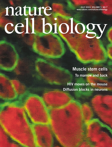
Muscle cells differentiate into haematopoietic lineages but retain myogenic potential. In the image, donor-derived bone-marrow cells differentiate into myofibres after injection into muscle. Dystrophin is shown in green and myosin heavy chain is shown in red. Cover design: Lawrence Keogh
E2F transcription factors coordinate the timely expression of genes during early cell-cycle progression. In addition, the E2F-1 subunit can induce apoptosis in response to DNA damage. New results reveal an unexpected function for E2F-1 in suppressing apoptosis, which may be important in explaining the contribution of E2F-1 to tumorigenesis.
Proteins that control the cell cycle would seem to have little to do with cell migration. However, a recent report indicates that Cdc2, a key cyclin-dependent kinase, can also enhance cell migration. Levels of Cdc2 are upregulated in cells that express high levels of the αvβ3 integrin, a protein that has long been implicated in enhanced migration and invasion of tumour cells.
Neurons exhibit distinct compositions in the axonal and dendrite plasma membrane, but it remains ambiguous whether or not a diffusion barrier is needed to keep the different components separated. Now, Nakada et al. utilize state of the art microscopy to follow the dynamics of single lipids or proteins inserted in different areas along the axonal and dendritic surface of neurons at different developmental stages. The results obtained shed new light on the mechanism underlying polarized segregation of membrane components in neurons.
Chordate claudins are core components of tight junctions. By contrast, VAB-9, a nematode four-pass transmembrane protein related to claudins, localizes to adherens junctions and contributes to cell adhesion and actin–plasma membrane association.