
Thank you for visiting nature.com. You are using a browser version with limited support for CSS. To obtain the best experience, we recommend you use a more up to date browser (or turn off compatibility mode in Internet Explorer). In the meantime, to ensure continued support, we are displaying the site without styles and JavaScript.

Volume 2 Issue 8, August 2000

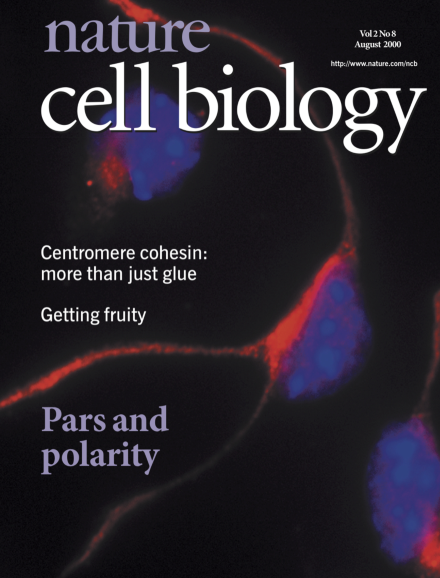
Deconvolution image of endogenous mPAR-6 in dissociated murine cortical neurons isolated from 16.5-day-old embryos. Eight hours after dissociation and plating, cortical neurons were fixed and stained with a polyclonal antibody raised against mPAR-6 (red). Neurons were counterstained with Heochst 333298 (blue). Endogenous mPAR-6 is enriched in perinuclear structures and filamentous structures in developing growth cones.

Article

Top of page ⤴

Brief Communication

Top of page ⤴

Editorial

Top of page ⤴

News & Views

Top of page ⤴

Book Review

Top of page ⤴

Historical Perspective

Top of page ⤴

Review Article

Top of page ⤴

Search

Quick links