

Key Points
- Achalasia is a syndrome of esophageal dysmotility characterized by failure of relaxation of the lower esophageal sphincter (LES) and lack of functional peristalsis in the body.
- All current forms of treatment for achalasia are palliative in nature.
- Pharmacologic therapy generally does not produce satisfactory or long-lasting relief.
- Other nonsurgical therapeutic choices include pneumatic dilation and botulinum toxin injection.
- Pneumatic dilation is one of the most well-established procedures for achalasia and is associated with a good initial response but only modest long-term improvement. About 50% of patients require a second or even third dilation in the first 5 years. There is a low but significant risk of perforation from this procedure.
- Botulinum toxin A also produces satisfactory initial results but its effects predictably wear off within a period of several months, necessitating further injections. It is generally safer and simpler than pneumatic dilation and hence may be valuable in patients at high-risk for the latter.
- Both pneumatic dilation and botulinum toxin do not work as well in younger patients.
- Diffuse esophageal spasm is related to achalasia and sometimes actually evolves into it. Diffuse esophageal spasm is often associated with LES dysfunction and responds to treatment directed at this sphincter such as pneumatic dilation, botulinum toxin, and surgery.
Introduction
Achalasia is a syndrome of esophageal dysmotility characterized by failure of relaxation of the lower esophageal sphincter (LES) and lack of functional peristalsis in the body. The vast majority of cases is acquired and probably represent a form of neurodegenerative disease of the enteric nervous system within the upper gastrointestinal tract. Clinical symptoms include dysphagia, chest pain, regurgitation, heartburn, and weight loss. It must be recognized at the onset that all current treatment for this condition is essentially palliative and is intended to overcome the resistance to esophageal emptying that is posed by the dysfunctional LES. Nevertheless, it is also true that in the vast majority of patients, satisfactory amelioration is eminently achievable with available therapeutic options.
Currently, all therapeutic approaches target the LES, with the specific goal of lowering resting pressure. This is because LES dysfunction is the cardinal feature of achalasia, leading to functional obstruction of the esophagus and responsible for most of the symptoms. Aperistalsis of the body of the esophagus, although required for the diagnosis, is generally considered as making a relatively minor contribution to the morbidity from this disease. Reduction in LES pressure can be achieved either by traditional smooth muscle relaxants, chemical denervation (i.e., botulinum toxin), brusque (usually pneumatic) dilation, or surgical myotomy.
Pharmacologic relaxation of the LES in the form of nitrates and calcium channel antagonists seldom translates into a satisfactory long-term strategy for the treatment of achalasia. In general, most patients tend to opt for other, more satisfactory forms of treatment after they have been on these drugs for a few months. This is because of incomplete relief, the need for multiple dosing, and significant side effects such as headaches, hypotension, and eventual tachyphylaxis. However, these drugs can be used on an as-needed basis pending investigations and during the planning stage for more definitive therapy.
Diffuse esophageal spasm (DES) is a less well characterized syndrome of spastic esophageal dysmotility than achalasia. Although the term (and more colorful variants such as corkscrew esophagus) has been used by radiologists for a long time, the current view is that it is best defined manometrically by the demonstration of intermittent simultaneous contractions in the body of the esophagus. The clinical and pathophysiologic aspects of this syndrome are discussed in detail in another section. However, some aspects are worth emphasizing from a therapeutic perspective. When associated with dysphagia, it is important to look for associated dysfunction of the LES. Hypertension or varying degrees of incomplete relaxation of the LES are commonly seen, ranging from half to more than two thirds of patients, and may contribute to the pathogenesis of symptoms, particularly dysphagia.1, 2, 3, 4, 5 Although this may be detected by standard techniques, the use of sleeve manometry may be particularly helpful in this instance,6 and this is routinely done in all patients with this diagnosis. The common association with LES dysfunction has led to the view that DES (or at least a subset of it) may represent either a forme fruste or a precursor of classic achalasia.7, 8, 9 This theory is supported in part by the few published studies on pathophysiology or pharmacology suggesting a loss of inhibitory innervation, similar to that seen in achalasia.5, 10, 11, 12, 13 These observations also provide the basis for targeting the LES for relief of dysphagia in these patients.
The next few sections of this review focus on pneumatic dilation and botulinum toxin injection, followed at the end by a discussion of the relative merits of current therapies and strategies for treatment of achalasia, followed by treatment of diffuse esophageal spasm.
Pneumatic Dilation (PD)
Rationale
Dilation of the LES, with the theoretical intent of disrupting its fibers, has the longest tradition among all the existing therapies for achalasia. Although now achieved by modern plastic balloons, little has changed in principle since the classic description of a patient treated with a whale bone by Sir Thomas Willis more than 300 years ago. Nevertheless, the biologic basis for relief from this procedure has never been satisfactorily established since at least one animal study showed no histologic evidence of damage to muscle despite marked reductions in LES pressure.14 What is clear is that mild degrees of stretching, as accomplished by simple bougies, are not adequate, resulting in relief that is both partial and transient.15 This has best been demonstrated in a controlled study of chagasic patients assigned to treatment with either a Browne-McHardy pneumatic dilator or a bougie (44 to 55 French), with the latter group failing to show any improvement in LES pressures and at 1 year reported no change or worsening of symptoms as compared with clear improvement in the former group.16 Currently, most gastroenterologists use a dilation of at least 30 mm to achieve satisfactory results. The specific device used to achieve this has evolved over the decades, from the mechanical Starck dilator, to hydrostatic and finally, pneumatic balloons.
Dilation Regimens
At the present time, the vast majority of dilations are performed using a plastic pneumatically inflated low-compliance 10-cm-long cylindric balloon (Rigiflex, Boston Scientific, Boston, MA) on a double-lumen catheter (one lumen for the balloon and the other for the guidewire). Three sizes of balloons, 30, 35, and 40 mm, are available. Less commonly, the Witzel dilator (American Endoscopy, Mentor, OH) is used17, 18, 19, 20; this consists of a 15-mm-long balloon mounted on an endoscope that is inflated under direct visualization and therefore does not require fluoroscopy. However, only one size of balloon, 40 mm, is available. The primary goal of any treatment for achalasia should be satisfactory improvement in symptoms. Nevertheless, because long-term outcomes vary considerably, a great deal of effort has been spent in figuring out optimal regimens including the number of dilations, diameter of balloons, duration of dilation, etc. In general, the standard ("graded") regimen consists of using a single dilator size at any one session (typically beginning with 30 mm); the symptomatic response over the next few weeks then determines the need for further dilations.21, 22 If this is deemed necessary, the next larger size is used and so on until the largest balloon has been deployed. A less popular regimen (the progressive method), consists of a series of progressively bigger dilations on the same or successive days, until "satisfactory" manometric or radiographic criteria are met.23 Although intuitively appealing, this method has never been directly compared and it is not clear whether in fact it leads to better results than the more conservative graded method.
Dilation Techniques
Dilation techniques vary widely, but there is broad consensus on common principles.22, 24, 25 The procedure is performed in a fluoroscopy suite and with the patient under conscious sedation. After a 12-hour fast (patients with severe retention may also need to be on a liquid diet for 1 to 2 days prior to the procedure), a careful diagnostic endoscopy is performed to assess the anatomy of the gastroesophageal junction, the cardia, and any other associated lesions. A guidewire [the stiffer Savary is preferred, although many investigators use a regular endoscopic retrograde cholangiopancreatography (ERCP)-type 0.035" wire] is then passed down into the distal stomach, its position noted by fluoroscopy, and the scope is withdrawn to the gastroesophageal junction, while at the same time keeping the wire in place. A marker is then placed on the endoscope at the mouth to indicate the approximate distance to the LES. The endoscope is then removed while maintaining the wire in its position. The distance to the LES (as noted on the scope previously) from the middle of the balloon is then also marked off on the catheter. The balloon catheter is then threaded over the guidewire until the desired length is inserted, typically with the middle of the balloon occupying the region of the cardiophrenic junction on fluoroscopy. A small amount of dilute radiographic contrast material may be injected into the balloon to assist in visualization; thereafter, under fluoroscopy, the balloon is gradually inflated with air with a pneumatic pump or syringe connected to a manometer, noting the position of the developing waist and making small adjustments to ensure that it occupies the center of the balloon for best results. Subsequently, the balloon is fully inflated; this usually requires about 120 mL of air at a pressure ranging between 7 and 15 psi. The balloon is kept inflated for 60 seconds; at this time patient discomfort may be severe, justifying the use of supplemental administration of narcotics immediately prior to full inflation. After 60 seconds, the balloon is rapidly deflated. Our practice is to repeat a full inflation for another 60 seconds, usually requiring a significantly less pressure than the initial dilation. At this time the deflated balloon and guidewire are removed and the patient transferred to the recovery area. The patient is observed for the next 5 to 6 hours during which serious complications, such as perforation, have usually declared themselves.26, 27, 28 We do not routinely do a contrast radiograph after the procedure. However, patients need to be monitored carefully while recovering, and promptly investigated in the presence of chest pain or other symptoms indicating a perforation. Otherwise, patients are discharged.
As mentioned, significant variations in technique exist among experts, with little or no evidence to suggest any effect on outcomes.21, 29, 30 These variations include the following:
- The rate, pressure, and duration of the initial dilation
- The need for a second dilation (or more)
- The need for a routine postdilation esophagogram
Clinical Results and Outcomes
Although PD has for years been regarded as the nonsurgical treatment of choice for most patients, a careful review of the literature reveals that it is far from perfect. The numerous publications attesting to its efficacy are retrospective in nature. As an example, two large reports of this kind are cited. In the first, a 65% success rate was reported in 899 patients with a mean follow-up of 6.5 years.30 In the second, a single PD was effective in 85% of 144 patients followed for an average of 6.5 years.31 Systematic reviews of these and other published studies suggest that about two thirds of patients have good (minor symptoms not requiring further treatment) to excellent (complete symptom resolution) improvement after one or more dilations in the first 5 years or so.32, 33
Although complete symptom resolution has been described in some patients followed for as long as 10 to 25 years,26, 31, 34 it is important to bear in mind that many studies include patients with short follow-ups, and further, many of the nonresponders may not have been included in the reported outcomes, having been lost to follow-up. Indeed, it has been shown that up to 40% of patients who do not respond may not seek medical help.35 Thus, it is important to analyze the results of the few prospective long-term studies that are available, which generally suggest a far less optimistic outcome following a single PD.36, 37 In an example of such a study, 54 consecutive patients treated with PD were followed every 2 years.37 Clinical remission was noted after a single dilatation in only 59% of patients after the first year, and 26% at 5 years. A follow-up publication on this same cohort (prolonging the observation period to a maximum of 19 years and obtaining a median follow-up of 13.8 years) showed a 5-year remission rate of 40% and a 10-year remission rate of 36%; repeated dilations only mildly improved the clinical response.38 Other studies have also shown that symptoms recur with time in up to 50% or more of patients,35, 39 with median dysphagia-free intervals of about 5 years.40
Pneumatic dilation may also not be equally effective for relieving all symptoms of achalasia. In one report, for example, PD had little effect on chest pain, which is present in approximately 40% to 60% of patients with achalasia.41
Predictors of Outcome
The two best predictors of outcome following PD appear to be postdilatation LES pressure and age.34, 37, 39 In keeping with the surgical literature, posttreatment LES pressures of 10 mmHg are associated with a better outcome; patients who achieved a postdilation pressure of <10 mmHg were much more likely to be in remission during follow-up compared to those with higher LES pressures (100% versus 23%).37 Younger patients (<40 years of age) also appear to do less well in response to pneumatic dilatation.37, 42, 43, 44 In the series discussed above, for example, the remission rate in patients older than 40 was much higher than in younger patients (67% versus 29%).37
Why do younger patients not do well? Although we do not know the answer, one can speculate on an anatomic rather than pathophysiologic reason. The LES appears to have two components with distinct anatomic, physiologic, and pharmacologic characteristics: (1) the classical ring-like or clasp muscle at the gastroesophageal junction, and (2) a sling of gastric fibers below this area45, 46, 47, 48, 49, 50, 51, 52 (Figure 1). The importance of the second component was shown in patients with achalasia who underwent separate division of the esophageal and gastric muscular fibers53; despite complete myotomy of the clasp muscle, significant tone was still maintained by the gastric sling fibers, the degree of which varied among individuals. Thus it is possible that these fibers contribute a major if not predominant tone to the LES in younger patients. These are not targeted by PD (or botulinum toxin), providing a possible explanation for the failure of endoscopic treatments in this subset of patients. Conversely, the better results with surgery may reflect the fact that the sling fibers are included in the myotomy.
Figure 1: Lower esophageal sphincter (LES) fibers.
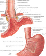
The two components of the LES are the horizontally oriented clasp fibers and the more vertically oriented sling fibers. (Source: Netter medical illustration with permission from Elsevier. All rights reserved.)
A role for male gender has also been reported as a possible factor determining outcome after PD.40, 54 In this regard, a number of other factors, such as duration of symptoms, the results of initial manometric parameters, nuclear emptying scintigraphy, and postdilation barium esophagrams have also been suggested.21, 34, 54, 55, 56 However, there is no definite evidence that any one of these is a reliable and/or independent predictor.
Complications
Pneumatic dilation is not an innocuous procedure, although it is relatively safe in expert hands. The most feared complication is perforation, reported in the literature in approximately 3% to 5% of patients in most series, with a range of 0% to 21%.15, 57 It is not possible to identify the patient at risk for perforation based on demographic, anatomic, physiologic, or technical factors.58, 59 Approximately 15% of patients complain of severe postprocedural chest pain,26, 42 but only a small proportion will actually be found to have a true perforation. Most perforations in this setting are small and may potentially be managed conservatively.58, 60, 61 However, unless the patient is at high risk for surgery, most centers will advocate prompt intervention and repair. The role for minimally invasive approaches such as endoscopic or videoscopic repair has yet to be defined.62, 63 Other traumatic complications of PD include tears, hematomas, and possibly diverticular formation.58
Gastroesophageal reflux disease (GERD) is uncommon after pneumatic dilatation, occurring in approximately 2% of patients overall.15 The likelihood of reflux probably correlates well with the efficacy of the procedure, and clinical monitoring for GERD symptoms should be part of routine follow-up.
Special Situations
Children
Pneumatic dilation has been performed in children as young as 2 years.64 Although earlier studies suggested that PD did not result in satisfactory response rates in children with achalasia, more recent reports suggest an outcome comparable to that of adults.64 Corresponding to their size, a smaller balloon, typically 20 mm in size may be used to begin dilations in children.
Pneumatic Dilation After Failed Previous Treatment with Surgery or Botulinum Toxin Injection
Pneumatic dilation appears to be safe in patients who have previously received botulinum toxin injection, with one report even suggesting an improved outcome in patients who had undergone PD after botulinum toxin injection, as compared with those who underwent PD alone.65 On the other hand, patients who fail surgical myotomy appear to do less well after PD as compared with untreated cases, with a symptomatic response seen in only about 50%, even though the risk of complications is no greater.66
Botulinum Toxin
Rationale
Botulinum toxin A (BoT-A) is one of the most potent inhibitors of acetylcholine release from nerve terminals, a property that has been exploited therapeutically in a wide variety of settings and more recently even in the gastrointestinal tract.67 The light chain of the toxin is a zinc endopeptidase that cleaves SNAP-25, a "snare" protein critical for the process of vesicular exocytosis.68 This results in a flaccid paralysis of skeletal muscle in the region of toxin injection. In the early 1990s, the toxin was also shown to reduce enteric smooth muscle tone, and since then it has been used for diseases such as achalasia, chronic anal fissure, sphincter of Oddi dysfunction, and gastroparesis.67, 69, 70, 71, 72, 73, 74 The rationale for its use in achalasia is based on the current pathophysiologic paradigm that there is a relatively selective loss of myenteric inhibitory neurons, resulting in unopposed excitation of the LES.75, 76, 77, 78, 79 By blocking the release of acetylcholine (and perhaps other excitatory neurotransmitters), BoT-A can attenuate their effects on LES tone.
Procedure
Botulinum toxin A is available commercially in the form of a lyophilized powder in vials of 100 units. Botox (Allergan, Irvine, CA) is the proprietary form of BoT-A available in the United States; 100 U are equivalent to 250 U of Dysport (Ipsen, Milan, Italy), available in other countries.80 It is reconstituted immediately prior to administration, typically to a concentration of 20 units/mL. For achalasia, the most common dose is 80 to 100 units, injected in 1-mL aliquots (20 to 25 units BoT/mL) in each of four quadrants about 1 cm above the Z-line or endoscopically visible sphincter rosette using a sclerotherapy needle through the channel of an endoscope.
Variations in this technique include a "2  4" technique (the total dose is divided into eight injections: four quadrant injections done at two different levels within the LES region),81 injection from below the gastroesophageal junction,82 as well as using endoscopic ultrasound83 and manometry84 as guides to more precise injection. However, there is no evidence that this improves efficacy and may not be necessary as the toxin is capable of diffusion for a limited distance in tissue. In a small study, endoscopic ultrasound was used to assess the location of the toxin immediately after blind injection in five patients with chagasic achalasia: about 85% of the injections were found to be located inside the muscle layers and only about 15% of injection points were found in the submucosa. Further, even those patients receiving submucosal injections of BoT-A showed significant improvement of their symptoms over a 6-month period.85
A comparison of three different dosing regimens, 50 IU, 100 IU, and 200 IU of BoT-A showed similar short-term response rates; however, a regimen of 100 IU given twice in a 30-day period produced the best long-term results, with about 68% patients remaining in remission for more than 24 months.81
The procedure is no more demanding than a routine endoscopy, and apart from an occasional complaint of transient chest pain, patients tolerate it well. They can go home after they recover from sedation and are allowed to eat later in the day. Symptomatic improvement occurs gradually and usually peaks 1 to 3 days later, although this may be delayed even further in the rare patient.
Clinical Results and Outcomes
Several controlled high-quality studies attest to the short-term efficacy of BoT-A for the palliation of symptoms in achalasia, with 1- and 6-month improvements seen in 75% to 100% and 44% to 100% of patients, respectively.67 The vast majority of studies also report an associated improvement in objective parameters, with the most commonly reported being a reduction in resting LES pressure, generally averaging around 40%.36, 71, 86, 87, 88 Although less commonly reported, radiographic or scintigraphic measures also improve.71, 89, 90, 91
Most of the published literature describes the experience with single, one-time-only injections; over time the effects of the toxin will wear off and neuromuscular communication is reestablished. Most, if not all, patients, therefore, will be expected to report recurrence of symptoms unless reinjected typically within 6 to 12 months of the first treatment, although some series have reported surprisingly long response durations averaging between 11 to 15 months or even longer.90, 92, 93, 94 It is clear, therefore, that repeated injections of BoT-A on an as-needed basis are required in order to maintain patients in remission. However, there are few studies that have evaluated the effectiveness of this strategy. In some series a trend toward a longer interval of response91 after the second injection has been noted, whereas others have reported no difference92 or the opposite effect.93 One study has approached this problem using a survival analysis method. Of those treated solely with BoT-A injections, repeated as needed, only 46% required pneumatic dilation or surgery by 2 years.95
Predictors of Response
For unclear reasons, up to a third of patients do not show a sustained response to BoT-A. This could be due to the fact that the LES in these patients is completely denervated and its tone is being generated by purely myogenic mechanisms. Alternatively, it may be due to the failure to target the "sling" component of the LES, similar to the situation described for pneumatic dilation (see above). Regardless of the mechanism, it is clear that the response rate is better (by nearly twofold) in older patients (e.g., >50 years of age),81, 90, 92, 96 a finding also observed with pneumatic dilation (see above). It also appears that patients with vigorous achalasia, irrespective of their age, have a good response,81, 92 perhaps reflecting an earlier stage of the disease process.76
As mentioned earlier, one study suggested that the dose of BoT-A and dosing schedule may also predict response.81 The study included 118 patients who were randomly assigned to receive one of three doses of BoT-A (50, 100, or 200 U). Patients randomized to receive 100 U were reinjected with an additional 100 U after 30 days at the end of follow-up. After 12 months, patients who received the two-dose schedule of 100 U were more likely to be in remission (80% versus approximately 55% for the other two groups).
Other factors such as duration of illness, previous dilation, or other forms of treatment (including incomplete myotomy), and baseline radiologic characteristics (including tortuous esophagus and epiphrenic diverticulum) may not influence outcome.91, 92, 97, 98
Complications
After a decade of use, BoT-A injections for achalasia can generally be considered safe. The low dose of toxin used for treatment of achalasia has virtually no risk of causing generalized neuromuscular blockade or paralysis. Immediate postprocedural transient and minor chest pain is reported in up to 25%, and heartburn may occur in up to 5% of patients.71
Esophageal wall injury and paraesophageal tissue inflammation have only rarely been reported clinically,99 and no significant mucosal or submucosal changes have been seen during endoscopic ultrasound following treatment.100 However, both PD and BoT-A injections caused similar degrees of inflammation and fibrosis in the swine esophagus.101 The histologic changes included acute and chronic inflammation with areas of increased fibrosis in the muscle. This is of potential concern because of possible interference with subsequent surgery, if required. Some surgeons have reported greater difficulty in performing myotomy after prior BoT-A injections, although this has been refuted by others.102, 103, 104 In another study that included 15 patients who had received preoperative BoT-A injections,105 difficulties in dissection of the submucosal plane were encountered significantly more often in the BoT-A group (53% versus 7%). However, there was no difference in postoperative symptom improvement between the two groups, and the operation was considered equally successful in both groups.
Although neutralizing antibodies have been detected in 10% to 20% of patients treated chronically with BoT-A for skeletal muscle conditions106, 107 their clinical significance remains unclear. It is not known whether this phenomenon occurs in patients with achalasia treated with multiple injections, and if it does, whether it could limit the efficacy of this approach.
Comparative Studies
Three methods are effective for treating achalasia: pneumatic dilation, botulinum toxin injection, and surgery. Pneumatic dilation has been compared to surgery for several decades. Most data seem to suggest that in expert hands, surgery offers a better chance of long-term relief, in keeping with the classic randomized trial reported by Csendes34 and summarized in Table 1. Several randomized trials have also attempted to compare these two modalities, as shown in Table 2. As can be seen, the 1-month clinical or manometric response to BoT-A is comparable to that to PD in four of the five studies. The response rate 1 year after a single injection is markedly inferior for BoT-A, which is to be expected given its pharmacologic properties, and as mentioned, repeated therapy with BoT-A is necessary to maintain efficacy.

Botulinum toxin A has also been compared with laparoscopic Heller myotomy; at 6 months, similar efficacy was seen.108 However, as expected, the probability of being symptom-free at 2 years was higher after surgery compared with after a single injection of BoT-A (88% versus 34%).
Quality of life after laparoscopic Heller myotomy with partial fundoplication, thoracoscopic Heller myotomy, PD, and BoT-A have been compared using a decision analysis.109 After 10 years, laparoscopic Heller myotomy with partial fundoplication was associated with the longest quality-adjusted survival. However, the absolute gain was small: 18 more days than pneumatic dilatation, which in turn was associated with 11 more days than BoT-A injections. These very small differences suggest that each approach is effective in terms of effectiveness in the initial management of achalasia. Further, these outcomes were sensitive to some practical variables such as the efficacy of laparoscopic surgery (to maintain its advantage, this needed to be more than 90%). On the other hand, BoT-A was superior to PD and laparoscopic surgery if prompt reinjection was performed after the recurrence of symptoms.
These regimens have also been compared on the basis of cost-effectiveness. A report using decision analysis found that surgical myotomy was the most costly strategy (costing approximately $10,800 compared to $3,245 and $3,911 for PD and BoT-A, respectively), despite higher short- and long-term efficacy.110 Pneumatic dilation was less costly than BoT-A so long as the rates of PD efficacy and perforation were >70% and <10%, respectively, and the cost of BoT-A (including endoscopy) was higher than $450. A Canadian cost-minimization analysis comparing BoT-A to PD suggested that BoT-A injections were less costly only if the life expectancy was less than 2 years.111 In a separately published economic analysis of the comparative trial between BoT-A and myotomy described above,108 the authors report that the initial cost was lower for Botox (1245 Euros) than for surgery (3555 Euros). However, at 2 years, this difference nearly disappeared: Botox 3364 Euros, surgery 3950 Euros.112
Strategies for Treatment
As a general rule, short-term results for both a single PD and one or two BoT-A injections are similar. Data regarding long-term outcomes are sparse for PD, whereas the long-term outcome after BoT-A injections is unknown. About 50% of patients with PD require further treatment at 5 years, whereas almost all patients treated with BoT-A relapse after a single injection eventually, usually in the first year or so. Surgical myotomy may provide a more permanent solution, but long-term reflux and its sequelae have to be taken into account.
Which treatment is best? Given the palliative nature of all treatments and the considerable variation in morbidity from the procedure, there is no simple answer to this question. One can, however, approach this issue from several perspectives. The first is based on direct head-to-head comparisons in the form of prospective trials, of which there are few in number. The second is an approach that relies on economic analysis of costs and effectiveness. Finally, and most importantly perhaps, the "right" therapeutic approach is based on the individual patient, taking into account his or her expectations, threshold for risk, and comorbidities.
This author, for instance, considers that a laparoscopic myotomy done by an experienced surgeon offers the single most permanent method of treatment for young adults. It should be emphasized, however, that aggressive monitoring and treatment of reflux is essential for long-term success. For otherwise healthy patients over 40 to 50 years of age or younger patients who are averse to surgery, pneumatic dilation is a reasonable first option, provided that they understand that there is a significant probability (up to 50% or even more) of requiring another treatment over the next 5 years. Finally, botulinum toxin can be considered for those patients who wish to "temporize" while making up their minds on a more permanent solution, those in whom the diagnosis is not clear cut113 those with serious comorbidities, or those who are averse to either surgery or pneumatic dilation. Whatever the method chosen, regular follow-up and in the case of surgery, monitoring of reflux, is imperative because the consequences of a neglected obstructed esophagus can be devastating and eventually lead to an esophagectomy.
Management of Diffuse Esophageal Spasm
Diffuse esophageal spasm is a relatively rare disease, and few clinical studies are available. Ideally, treatment DES should be able to eradicate the symptoms and restore esophageal propulsive peristalsis. Currently we are far from achieving these goals with any of the available treatments. Surrogate goals include alleviating the major symptoms of dysphagia, noncardiac chest pain, and regurgitation as well as improving the esophageal bolus transit.
Medical Treatment of Diffuse Esophageal Spasm
There are only few reports on clinical trials in DES. Anticholinergics (propantheline, cimetropium, ipratropium), nitrates, and calcium channel antagonists yielded inconsistent results.114, 115, 116, 117, 118 The calcium channel antagonists nifedipine and diltiazem failed to demonstrate significant benefit over placebo in patients with DES in double-blind controlled studies.118, 119 The  -adrenoreceptor salbutamol, which was reported to be effective in a single case, may become a novel choice in the treatment of patients with DES.120
Botulinum Toxin (BoT-A) Injection
The effect of BoT-A injection has been described in the discussion of achalasia treatment. Likewise, BoT-A has been employed in the treatment of DES for several years. At present, there is no double-blind randomized study investigating the effect of this therapeutic option. Few relatively small studies reported good clinical results in short- and long-term alleviation of symptoms secondary to DES. At least two reports, both of uncontrolled trials, suggest a 70% to 80% improvement in symptoms after BoT-A injection in the LES as well as in the body.121, 122 Further, initial responders did well after second injections for recurrent symptoms.
Pneumatic Dilatation
Similarly to reports on surgical myotomy in this syndrome,123, 124, 125 it is logical to presume that endoscopic therapies aimed at the sphincter may also be successful. Although the literature is much too sparse to make firm recommendations, it appears that pneumatic dilation of the LES126, 127, 128 or occasionally of only the esophageal body129 may be beneficial in this syndrome. As an example, a retrospective study using a hydrostatic dilator reported a significant improvement in dysphagia in eight of nine patients with DES and LES dysfunction followed for an average of about 3 years.130 These patients had previously been unresponsive to medical therapy and simple bougienage. Other reports have been less positive, showing only a 45% improvement.23
In conclusion, a case can be made for a vigorous search for LES dysfunction in patients presenting with DES, and if found, to treat these patients as if they were to have achalasia. It should be noted that dysphagia is the symptom most likely to respond to such an approach, and other symptoms such as chest pain may have to be treated with pharmacotherapy including the use of neuropathic analgesics such as tricyclic antidepressants.
Surgical Treatment of Diffuse Esophageal Spasm
Surgery is considered a last resort for many reasons. First, there are no double-blind randomized studies in this area attesting unequivocally to the efficacy of this procedure. Further, published reports often deal with global results in heterogeneous groups of patients with no distinction being made between diffuse esophageal spasm, nutcracker esophagus, hypertensive LES, achalasia, and sometimes even anatomic abnormalities (diverticula).131, 132, 133, 134 Most patients presenting for surgery have a longstanding history of symptoms, although in some cases conservative treatment was abandoned in as brief a period as 1 year.133, 135 Currently there are several surgical options considered in the treatment of diffuse esophageal spasm. There is no good scientific basis for what is currently considered the procedure of choice: long esophageal myotomy. The proximal limit of myotomy as well as the inclusion of the LES continues to be an area of disagreement among surgeons.133, 134, 136 As with achalasia, laparoscopic and thoracoscopic approaches offer some advantages and are becoming increasingly popular.131, 132, 137 When myotomy includes the LES, as is usually the case, an antireflux procedure should be included.131, 133, 135, 137
Open myotomy has been reported to improve symptoms in 70% to 90% of cases,123, 136, 138 a rate matched or exceeded by less invasive surgical approaches (80% to 94%).131, 132, 134, 137 Not all the published results are as favorable, however, particularly in the long-term. In a median of 6 years' follow-up in eight patients who underwent open esophageal and LES myotomy for "spastic disorders" of the esophagus, only three patients remained symptom free. In another four patients, esophagectomy was finally offered for progressive esophageal dilatation and dysphagia.133 Both procedures are generally considered safe with little or no mortality.131, 132, 133, 134, 135, 139 Late complications included dysphagia or chest pain in 28.5% and GERD in 14.2%.131
