
Figure 2 - Brainstem swallowing sites and activity of swallowing neurons.
From the following article
Electrophysiologic characterization of the swallowing pattern generator in the brainstem
André Jean and Michel Dallaporta
GI Motility online (2006)
doi:10.1038/gimo9
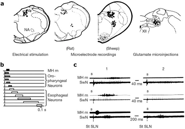
a: Drawings of coronal hemisections of the brainstem, at the level of the intermediate subpostremal part of the nucleus tractus solitarii (NTS), in rat and sheep. Dots indicate the localization of active points, stimulation of which induces swallowing, of swallowing neurons in rat or sheep (microelectrode recordings), and of glutamate injection sites inducing deglutition. b: Diagram showing the typical swallowing sequence of different medullary NTS interneurons [vertical lines, superior laryngeal nerve (SLN) stimulation; black rectangle, mylohyoideus contraction (MH m); gray and white rectangles, discharge of oropharyngeal or esophageal neurons]. c: Oropharyngeal and esophageal neurons (SwN) recorded before (1) and after (2) motor paralysis (gallamine 2 mg/kg). Note that the neuronal discharge remained unaltered after motor paralysis. (Source: Adapted from Jean.6, 109 Jean et al. 110, Kessler and Jean 21, Kessler et al. 27. with permission )
Powerpoint slides for teaching
If the slide opens in your browser, Select "File > Save as" to save it.
Download Power Point slide (738K)