Figures, tables and video
From the following article
Surgical treatment for achalasia
Jedediah A. Kaufman, Dave R. Lal and Brant K. Oelschlager
GI Motility online (2006)
doi:10.1038/gimo53
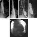
Figure 1
Esophagrams of a patient with early achalasia pre- and posttreatment.
Full size figure and legend (47K)