
Figures, tables and video
From the following article
Sachin Wani and Prateek Sharma
GI Motility online (2006)
doi:10.1038/gimo45
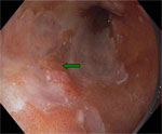
Figure 1
Endoscopic image of Barrett's adenocarcinoma using high-resolution endoscopy.
Full size figure and legend (13K)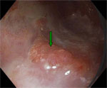
Figure 2
Closer view of the nodular mucosa of Barrett's adenocarcinoma (green arrow) using high-resolution endoscopy
Full size figure and legend (12K)
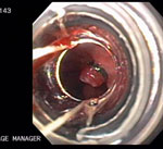
Figure 5
Endoscopic mucosal resection of early Barrett's adenocarcinoma using the banding technique.
Full size figure and legend (63K)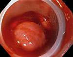
Figure 6
Barrett's adenocarcinoma specimen in situ post–endoscopic mucosal resection.
Full size figure and legend (58K)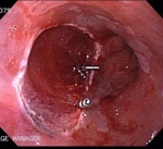
Figure 7
Endoscopic picture of the distal esophagus post–endoscopic mucosal resection.
Full size figure and legend (65K)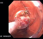
Figure 8
Specimen retrieval post–endoscopic mucosal resection using a net basket.
Full size figure and legend (54K)
Table 2
Staging of esophageal cancer according to the American Joint Commission on Cancer system
Full size table and legend