
Figure 12 - An in vitro model of esophagitis.
From the following article
Signal transduction in lower esophageal sphincter circular muscle
Piero Biancani and Karen M. Harnett
GI Motility online (2006)
doi:10.1038/gimo24
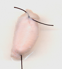
The mucosa/submucosa is freed from esophageal muscle, by sharp dissection at the level of the submucosa. Segments of the mucosal tube are tied at both ends, forming a closed sac with the squamous epithelium on the inside and the submucosa on the outside. The sac with HCl (pH 5.8) or Krebs buffer inside, is kept in oxygenated Krebs buffer supernatant for 3 hours (1 mL Krebs buffer/100 mg mucosa). In response to HCl, the mucosal sac produces and releases inflammatory mediators into the supernatant, which may then be applied to muscle strips, or used for measurement of inflammatory mediators. (Source: Cheng et al.,105 with permission from the American Physiological Society.)
Powerpoint slides for teaching
If the slide opens in your browser, Select "File > Save as" to save it.
Download Power Point slide (1,836K)