Sir,
Complications associated with alloplastic implants (silicone/silastic/supramid) are rare.1 However, they can occur some considerable time after surgery.2 A 36-year-old Caucasian male presented with complete lower eyelid retraction 3 years after silicone sheet orbital floor implant. The implant extruded spontaneously 3 days prior to eyelid reconstructive surgery and was not replaced. The surgical technique is discussed, and a brief discussion of the complications of alloplastic implants presented. More modern porous implants may reduce the chance of implant extrusion and lower eyelid retraction.
Case report
The patient initially presented to our unit with a left orbital cellulitis. Three years previously he had undergone a left orbital floor fracture repair with insertion of a silastic implant at another hospital. On this occasion, he was treated with oral antibiotics and the infection subsided. However, he represented 3 months later with an unsightly red left eye. Examination revealed a complete retraction of the left lower lid, extensive chemosis of the inferior bulbar conjunctiva, and restriction of upgaze in the affected eye (Figure 1).
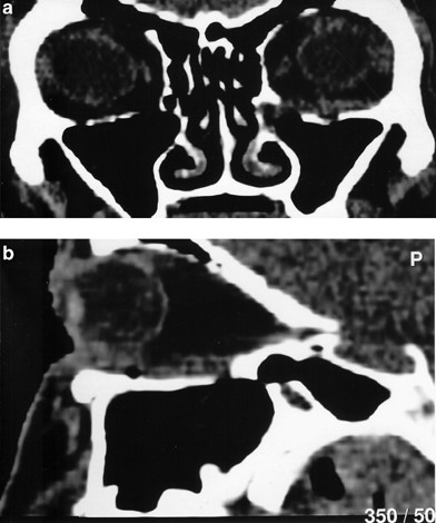
A CT scan of the orbits revealed a well-defined opacity in the region of the orbital floor implant (Figure 2a and b). The inferior rectus cannot be differentiated from this.
At 3 days prior to elective surgery, the silastic implant extruded spontaneously. Exploration revealed an opening in the inferior conjunctival fornix, through which the implant had presented. Reconstructive surgery was performed. The eyelid margin, tarsal plate, and anterior lamella of the lower eyelid had completely retracted along the floor of the orbit. A dense fibrous tissue reaction around the implant had caused this. The lower eyelid was carefully dissected from the deep fibrous tissue, with care not to damage the inferior rectus. The retractors were recessed and an abdominal dermis fat graft was harvested. This was placed along the inferior orbital margin. A hard palate graft was also harvested. This was sutured into place between the lower lid retractors and the lower border of the tarsus.
The orbital implant was not replaced. The immediate postoperative appearance is shown in Figure 3. The patient was instructed to follow an intensive regime of wound massage for 4 months. Figure 4 shows the appearance of the patient at 9 months postoperatively. He was pleased with the cosmetic result. Ocular motility was normal but there was residual limited lower lid retraction on downgaze.
Comment
Alloplastic implants can become encapsulated within a dense fibrous capsule, which can make treatment of implant infection very difficult.3 Occasionally, fibro-vascular tissue around nonintegrated implants can haemorrhage and cause acute swelling of the capsule. In such cases, patients present with pain and proptosis.2 The haematoma may spread beyond the capsule to compromize the orbital apex. Unless this is promptly recognized, blindness may result.
More recently, porous implants have been used. These allow better integration into the hosts' tissues. Fibrovascular proliferation occurs through the implant. Thus, helping to stabilize the latter and reduce the theoretical risk of migration. If an implant were to move anteriorly, this would cause direct pressure on the mucosa and subsequent extrusion or fistula formation may result.4, 5 An unstable implant can also move posteriorly towards the optic nerve.6, 7 Blindness may result from compression of the latter. For this reason, an implant should never be placed more posteriorly than the posterior wall of the maxillary sinus.
Other recognized complications from alloplastic implants include extraocular muscle entrapment,8 dacrocystitis,3, 9 presumably from local spread of infection, hyperglobus,10 and ectropion with scleral show from excessive scarring.10 Hyperglobus may result from implant stacking or the formation of granulation tissue around the implant. The formation of cysts has been reported in the use of gelatin film alloplastic grafts.11 These form when there is partial reabsorption of the gelatin implant into the tissues.
The treatment of all the complications is the same; remove or reposition the implant. In practice, the surgeon may find it difficult to identify normal structures due to extensive scar tissue and bleeding from fibrovascular sources. It is very important to visualize all margins of the fracture. All herniated orbital tissues must be repositioned. The orbital floor defect must be covered completely without orbital tissue herniation; the smallest implant that will achieve this in the subperiosteal position is ideal. The ideal material will induce little scarring or tissue reaction. The majority of ophthalmic surgeons use alloplastic implants. It is, therefore, worthwhile explaining to patients the potential complications of their use.
References
Jordan DR, St Onge P, Anderson RL, Patrinely JR, Jeffrey A, Nerad MD . Complications associated with alloplastic implants used in orbital floor fracture repair. Ophthalmology 1992; 99: 1600–1608.
Mauriello JA, Flanagan JC, Pegster RG . An unusual late complication of orbital floor fracture repair. Ophthalmology 1984; 91: 102–107.
Mauriello JA, Fiore PM, Kotch M . Dacryocystitis: late complication of orbital floor fracture repair with implant. Ophthalmology 1987; 94: 248–250.
Goldman RJ, Hessburg PC . Appraisal of 130 cases of orbital floor fracture. Am J Ophthalmol 1973; 76: 152–155.
Wolfe SA . Correction of lower eyelid deformity caused by multiple extrusions of alloplastic orbital floor implant. J Plastic Reconstr Surg 1981; 68: 429–432.
Burres SA, Cohn AM, Mathog RH . Repair of orbital blowout fractures with marlex mesh and gelafilm. Laryngoscope 1981; 91: 1881–1886.
Weintraub B, Cucin RL, Jacobs M . Extrusion of an infected orbital floor prosthesis after 15 years. Plastic Reconstr Surg 1981; 68: 586–587.
Mauriello JA . Inferior rectus muscle entrapment in teflon implant after orbital floor fracture repair. J Ophthalmic Plastic Reconstr Surg 1990; 6: 218–220.
Kohn R, Romano PE, Puklin JE . Lacrimal Obstruction after migration of orbital floor implant. Am J Ophthalmol 1976; 82: 934–936.
Browning CW . Alloplastic materials in orbital repair. Am J Ophthalmol 1967; 63: 955–962.
Lotfield K, Jordan DR, Fowler S, Anderson RL . Orbital cyst Formation associated with gelafilm use. Ophthalmic Plastic Reconstr Surg 1988; 3: 187–191.
Author information
Authors and Affiliations
Corresponding author
Rights and permissions
About this article
Cite this article
Vose, M., Maloof, A. & Leatherbarrow, B. Orbital floor fracture: an unusual late complication. Eye 20, 120–122 (2006). https://doi.org/10.1038/sj.eye.6701801
Published:
Issue Date:
DOI: https://doi.org/10.1038/sj.eye.6701801


