
Key Points
-
Identification and definition of the patient's aesthetic problem
-
Consideration of the balance between aesthetics and tooth destruction for conventional and adhesive restorations
-
An awareness of the aesthetic limitations of restorations and an attempt to ensure that the patient's expectations are realistic
-
Incorporation of procedures leading to better aesthetics at each clinical stage
-
Confidence in determining shade and communicating effectively with the laboratory
Key Points
Crowns and extra-coronal restorations:
-
1
Changing patterns and the need for quality
-
2
Materials considerations
-
3
Pre-operative assessment
-
4
Endodontic considerations
-
5
Jaw registration and articulator selection
-
6
Aesthetic control
-
7
Cores for teeth with vital pulps
-
8
Preparations for full veneer crowns
-
9
Provisional restorations
-
10
Impression materials and technique
-
11
Try-in and cementation of crowns
-
12
Porcelain veneers
-
13
Resin bonded metal restorations
Abstract
A pleasing dental appearance is the subjective appreciation of the shade, shape and arrangement of the teeth and their relationship to the gingiva, lips and facial features. Achieving such a pleasing appearance in our patients is not always easy but is critical, not least because our work is effectively on display and this has implications for patients' perceptions of our practice. To be successful, thorough assessment, careful planning and precise clinical execution is required. Every bit as important though, is good communication, both with the dental laboratory and particularly with the patient. In few areas of dentistry can effective communication be as critical as it is here.
Main
Retention of natural teeth into old age is now commonplace and whilst usually desirable, it has brought with it considerable additional problems. Making well-aligned white teeth in a complete denture is usually straightforward, but matching a single crown or veneer to a group of natural incisors is a different matter altogether. This problem is illustrated by data from the 1988 survey of adult dental health in the United Kingdom1which showed that having just one or two crowns was more likely to be associated with dissatisfaction with the appearance than having none or many.
In each case, planning tooth preparation involves the dentist in a cost:benefit analysis, where the cost of improved aesthetics is judged in terms of removal of tooth tissue and in the potential for damage to the pulp and periodontium. However the benefit of stunning porcelain work is easiest to achieve where a thick layer of material can be used to develop the optimum optical properties, but this usually requires more tooth tissue to be removed. This is a theme which runs right through all aesthetic considerations and should underpin what follows. This concept does not sit comfortably with a dogmatic approach with hard and fast rules about the dimensions of a preparation. The clinician will choose to alter the cost:benefit balance in different ways in different cases. The choice might be made to sacrifice aesthetics for long-term health, or to take a risk with long-term pulp health to maximise aesthetics; for example with a heavily prepared ceramo-metal restoration.
Aesthetic improvements are most important for anterior teeth and may often be the sole reason for providing the restoration. The type of materials used clearly have an important bearing on both the appearance and the amount of preparation and are an important part of the aesthetic cost:benefit equation. Table 1 lists the aesthetic restorations commonly available. Whilst they are much less destructive of tooth tissue than traditional ceramo-metal crowns, adhesive restorations such as the porcelain laminate veneer and dentine bonded crown do have limitations: specifically the problem of masking the colour of darkly stained teeth, problems of temporisation and the inability to cement restorations provisionally. Veneers are covered in detail in a separate article in this series (Part 12). Furthermore whilst there have been significant improvements in indirect composite technology there is as yet little clinical evidence of their stability and longevity.
The key decisions are similar for anterior or posterior teeth, but there is usually less room for aesthetic compromise at the front of the mouth. On posterior teeth it may be feasible to sacrifice optimum aesthetics by restricting the use of porcelain only to the most visible sites and consequently cutting a less damaging preparation. For example on a short tooth, creating space for occlusal porcelain and metal rather than for metal alone could make the difference between success and failure of retention. Furthermore, tooth preparation carries with it the risk of pulp damage.2
A conservative approach would equate not only to less pulp morbidity, but more tooth remaining should the need arise to remake the restoration. On a posterior tooth it may also be possible to use a three quarter crown, which leaves the bulk of the buccal surface intact. Once again, whatever the materials chosen, it is important that the patient fully understands the advantages and limitations of the restorative solution. This article aims to address all of these issues. The field of dental aesthetics is highly subjective and, as a result, difficult to research. The advice we offer and the recommendations we give are necessarily based more on experience than scientific analysis.
Identifying the problem
The first and fundamental key to obtaining a successful aesthetic result is to establish the precise nature of the patient's demands, at the outset of treatment. What is perceived as 'natural' or pleasing to the dentist or technician may be much too 'natural', and far from pleasing for the patient. This may sound obvious, but without a detailed assessment it is easy to fail to make a precise diagnosis of the patient's desires, and so end up treating something which is not a problem for the patient, or creating technically beautiful restorations which the patient perceives to be aesthetically unsatisfactory, with all the angst and cost that this implies.
Table 2 shows the many factors, which must be considered in defining the patient's aesthetic problem. On the basis of a thorough assessment, the dentist must decide whether:
-
The patient's expectations are realistic
-
The proposed treatment options are in the patient's best interests
-
The dentist, with the support of the laboratory, has the skill to carry out the treatment.
One of the greatest challenges for dentists providing crowns and veneers is the need to match expectations to what is technically and aesthetically achievable.
Matching expectations with reality
Most patients appreciate a full and frank discussion about what is achievable. Time spent at this stage can save a lot of heartache and expense later on. There are reversible means of helping patients understand the scope and possible consequences of treatment:
-
Wax mock-ups (diagnostic wax-ups) on stone casts can be very useful for demonstrating treatment options and act as blue-prints for carrying out clinical and laboratory work (Figs 1 and 2). Some dentists prefer these models to be created in tooth-coloured wax whilst others express concern that patients may not fully appreciate that these models are to assess shape not colour and for this reason deliberately use non tooth-coloured wax.
Figure 1 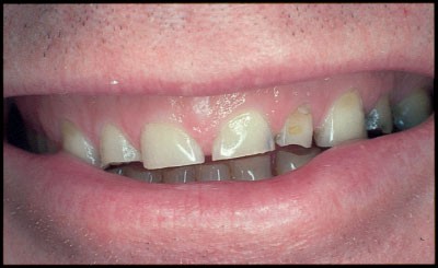
Short anterior teeth causing an aesthetic problem
Figure 2 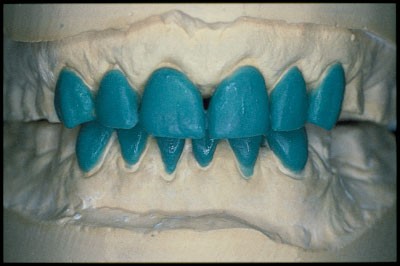
Diagnostic wax-up of Case in figure 1
-
Composite resin can be used on teeth in its uncured state to indicate the potential for shade and additive shape changes to natural teeth (Fig 3 to 5).
Figure 3 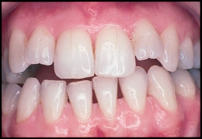
Case requiring changes in upper lateral incisor length and levelling of lower incisal plane
Figure 5 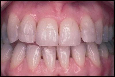
Final restoration of upper lateral incisor edges of case in figure 3 (directly applied composite resin used in this instance)
-
When viewed against the darkness of the mouth, black, water-soluble ink can give a rough idea of the effect of subtractive shape changes such as shortening overerupted lower incisors (Fig. 4).
Figure 4 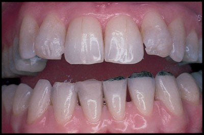
Trial alterations to case in figure 3: uncured composite added to upper incisal edges and water-soluble ink to lower left incisors and canine
-
Computer software, which can manipulate photographic images, is becoming commonplace in the dental surgery and can be useful in discussions of treatment options. However,it is probably fair to say that it is much more difficult to provide restorations exactly as created on a screen than to use a wax model as a guide, especially if the patient has been given a colour enhanced print of the expected outcome! Unlike the techniques described above, computer manipulation of images has no physical limits and may as a result create unrealistic expectations.
-
Photographs of previous cases may help patients to understand both the possibilities and the limitations. Restricting these to the ones with the best results may be a mistake. Where there are clearly going to be aesthetic limitations, it is probably best to illustrate them with realistic examples. The patient may be more likely to agree to treatment if they can picture the outcome, rather than imagine it from a verbal description.
-
Provisional restorations can allow the subtle relationship between the shape and form of the teeth and the soft tissues and facial features to be evaluated and decided before the final restoration is constructed. They can also ensure that tooth preparation for restorative material is adequate (by establishing the desired shape and form and then measuring the thickness of temporaries with callipers).


Unrealistic expectations
Some patients may demand changes in appearance which are objectively difficult to appreciate and still more difficult to realise. In most cases this is simply a problem of communication, but unrealistic expectations and a history of multiple previous treatments addressing appearance may be a warning of a patient with Body Dysmorphic Disorder (BDD) or Dysmorphophobia3: a preoccupation with a defect in appearance which is either imagined or excessive in relation to a minor defect and which causes significant distress in social, occupational and other areas of life. BDD is probably rare but is an extraordinarily difficult problem to deal with. It is unlikely that demands to change appearance will be satisfied for this group of patients. A second opinion is a perfectly acceptable course of action if in doubt.
Final planning and clinical procedures
Having decided on the restoration type, it remains to finalise margin features and carry out the clinical stages, ultimately leading to cementation.
Supra or sub-gingival: where should the crown margin go?
Factors identified in the assessment should help to determine the location of crown margins. Where the margins are not visible, there are good biological arguments for placing all margins supra-gingivally.4 These sites would include all margins on molars, lingual and interproximal sites, and buccal aspects of anteriors and premolars where functional lip positions obscure the gingival margin. Not only will this facilitate finishing and maintenance but should also favour periodontal health. The appearance of a supragingival margin can be optimised by ensuring that the finish line is in harmony with the level of the gingival margin.
Where exposed crown margins are likely to create an aesthetic problem, margin placement up to 1 mm into the gingival sulcus can be acceptable.5 However, great caution should be exercised as gingival margins are at risk of recession, particularly when there are prominent roots, thin gingival tissues or in the presence of periodontitis. Though periodontal attachment loss occurs to a greater or lesser degree through life,6 trauma during clinical stages and poorly fitting temporary and final restorations will increase the risk of gingival inflammation and subsequent recession. The need to optimise retention by increasing preparation length may be better addressed by removing periodontal attachment surgically ('crown lengthening'), than by making the margin encroach deeply into the gingival sulcus and attachment apparatus with the risk of inflammation.
Shoulder or chamfer: what should the preparation finish line be like?
There is a forceful argument that where possible, ceramo-metal crowns should have metal margins because this produces the most predictable marginal seal7 but as discussed earlier in this series (Part 2 'Materials Considerations'), this is a contentious issue. However by avoiding the metal collar, a porcelain butt fit, created on a shoulder finish line, will generally allow for better aesthetics in critical areas. A restoration whose margin is in porcelain may allow light to pass into porcelain from the gingival aspect as it does into intact teeth contributing to a lifelike appearance.8
How much metal: where should the porcelain-metal junction on ceramo-metal crowns be?
There is no biological or technical benefit in using porcelain at sites that are not visible. Consideration given to the precise location of porcelain-metal junctions for ceramo-metal crowns at the planning stage gives the potential to optimise conservation of tooth structure yet still maintain satisfactory aesthetics. Volume to volume, the extent of reduction for metal alone is substantially less than for metal and porcelain: different depths of tooth reduction can be used at different sites depending on the covering material(s). Tooth preparation then becomes an ordered technical exercise to satisfy the need for differential space attainment. It should be obvious to the technician examining the resulting die where to locate porcelain-metal junctions (Fig. 6). There are laboratory cost implications to provision of ceramo-metal crowns of this sort. It is necessary to wax a full contour restoration on the die, mark the porcelain-metal junction and then cut back space in the wax pattern for porcelain rather than simply to create a thin metal coping over the whole preparation which is covered by porcelain.

Die with reduction to match veneering material(s): the larger buccal axial reduction is needed to accommodate metal and porcelain, the smaller palatal reduction is for metal alone
Shade matching
Shade matching is something many of us find difficult and is often done last whereas in fact it should be done first! It is not an exact science, involving as it does a good deal of subjective judgement. Although an accurate reproduction of shade is an obvious goal, it cannot be divorced from consideration of shape, surface texture and special characteristics, which are described later. Teeth possess a range of optical features seemingly designed to make shade matching difficult! Teeth:
-
Are non-uniform in colour
-
May have complex visible internal and surface features
-
Are semi-translucent
-
Exhibit a degree of fluorescence
-
Change shade and shape with age
In addition, a good shade match to porcelain in one light condition may be a poor one under different lighting: a phenomenon termed metamerism. Despite these obstacles, the best porcelain restorations go a long way to reproducing nature using a combination of skilful artistry and optical trickery. Before recording and prescribing shade it is useful to have a basic understanding of the science and dimensions of colour and texture so that shades can be interpreted and communicated precisely.
Dimensions of colour
Colour can be described in terms of three dimensions:
-
1
Hue: The name of the colour eg blue, red etc.
-
2
Value: An achromatic measure of the lightness or darkness of a particular colour such that high value refers to a shade which is light and low value to one which is dark. Two completely different colours can have exactly the same value. To help understand this, imagine the effect of black and white television on colours.
-
3
Chroma: The strength or saturation of a colour of particular hue. Imagine increasing the chroma of a small amount of colour pigment diluted in water by adding more of the same pigment.
Shade guides in common use (Figs 7 and 8) are not designed for a systematic assessment of the dimensions of colour and have been criticised for not including a broad enough range of shades. Two commonly used guides (Vita Lumin and Ivoclar Chromoscope) are composed of groups based essentially on hue (Vita Lumin: A= reddish brown, B = reddish yellow, C = grey shades, D = reddish grey, Ivoclar Chromascop: 1 series = cream, 2 series = orange, 3 series = light brown, 4 series = grey, 5 series = dark brown), with sub-classes of varying value and chroma. The Vitapan 3D Master system uses a simple but methodical approach to shade determination based on the three dimensions of colour. In common with previous work,9 the Vitapan 3D Master system emphasises value as the most important dimension in colour matching for porcelain restorations. Whichever guide is used, it is useful to understand colour terminology as it forms a language for communicating additional information about colour to the laboratory.

Vita Lumin shade tabs with stained necks removed in order of decreasing value

Ivolcar Chromascop shade guide
Surface texture
This quality describes surface contour both at a 'macro' level, such as developmental lobes and ridges, as well as fine surface detail such as perikymata. The lustre of a restoration describes the level of glaze produced in the porcelain oven or by various rotary instruments and polishing techniques. Lustre can effect value perception such that high lustre raises value. It is therefore an important feature to match and one which is often neglected. At the very least, terms such as high, medium or low lustre can be used on the prescription, and are more effective if they are linked to a standardised reference guide which can be used both in the surgery and in the dental laboratory. The technician can often get a good indication of other surface features from surrounding teeth.
Special characteristics
These include fracture lines, white spots and translucency. The best looking special characteristics are incorporated during incremental porcelain application. Surface stains can be used to produce some of these effects but are prone to wearing away with time.
Choosing and prescribing a shade
As well as factors inherent in teeth themselves, barriers to accurate shade matching often include inappropriate viewing conditions, shade guides that tend to be made from thick layers of high fusing porcelain and even colour blindness.10 Against this background, it is perhaps not surprising that different dentists tend to match the same tooth differently from each other, and even from previous attempts to match the same tooth themselves.11 Choosing a shade will benefit from adherence to a protocol based on sound reasoning. Table 3 gives a method for assessing shade and surface texture12 which we think is highly appropriate and is easy to apply. Occasionally custom-made shade tabs with varying amounts of porcelains and a range of surface textures and special characteristics are helpful. Electronic optical devices have been produced to assist making an objective assessment of shade but their usefulness remains to be fully evaluated.
Tooth preparation
Achieving optimum aesthetics depends heavily on providing the technician with adequate space for the incremental application of porcelain (Fig. 9). The considerations have already been discussed above, but when it comes to the practicalities, the extent of tooth preparation is best visualised intra-orally by reference to a preparation guide. A small putty mould, made over the tooth before preparation, and then cut in cross-section is invaluable if the shape of the tooth is to be maintained. A putty mould (Figs 10 and 11) or vacuum formed matrix made from a diagnostic wax-up is required if the shape of the tooth is to be changed (Fig. 12). Depth cuts to guide tooth reduction may be a useful guide to ensure adequate reduction, but are not very helpful when shape changes are planned. Matrices are particularly helpful on the buccal surfaces of upper anterior teeth which are curved when viewed from the mesial or distal. There is a tendency to prepare the buccal surface in a single plane, ignoring the curvature (Fig. 13). The aesthetic result will either be a bulky crown (if the full thickness of porcelain is placed), or a crown where the contour is correct, but where the core porcelain is inadequately masked. To achieve a good aesthetic result the buccal surface preparation should follow the natural curvature of the tooth (see Fig. 11).

Space required for metal coping and layers of porcelain for a ceramo-metal crown

Putty mould sectioned and numbered on a diagnostic wax-up

Putty matrix in-situ to help visualise appropriate tooth reduction

Vacuum formed matrix in-situ

Crown preparation with no second plane of reduction
Clinical records
As well as the role of the facebow record in helping to make movements of casts on an articulator anatomical, it ensures that articulated working casts are orientated to the base of articulator in the same way that the patient's teeth are orientated with respect to the floor (if the patient's head is upright and the anatomical features used as reference points are normally related!). This helps the technician 'see' the restorations orientated as they would be when observing the patient. Very occasionally an ear-bow recording can give an erroneous interpretation of the relationship of the occlusal to the horizontal plane. This discrepancy occurs as a result of the patient's ears being at different levels and may need to be compensated for where multiple anterior crowns are prescribed.
Try-in and cementation
Where shade matching has been difficult for conventional crowns, there is merit in trial placement of moistened restorations before giving them their final surface finish. Surface stains and changes in surface form can be prescribed at this stage (Fig. 14). It should be remembered that surface stains might eventually be lost.

Along with written instructions, pencil marks help to indicate changes required
After glazing, a period of trial cementation leaves scope for a further period of assessment by both patient and dentist. If restorations are subsequently returned to the laboratory for adjustment, steps must be taken to dehydrate porcelain before firing to avoid the risk of fracture. Furthermore, temporarily cemented definitive crowns can be difficult to remove. Zinc oxide and eugenol-based cement must have its mechanical properties significantly reduced by adding a modifier as described later in this series (Part 11 'Try-in and cementation').
Pigmented luting agents allow subtle manipulation of shade for adhesive porcelain restorations. Some systems provide water-based trial cements to facilitate the choice of colour. Manufactures' instructions should be followed.
Communication with the laboratory
The dentist must accept ultimate responsibility for all aspects of completed laboratory work. On the face of it this might suggest that a totally prescriptive one-way communication is required. Not surprisingly such an attitude can lead to feelings of frustration and dissatisfaction to all concerned. It does not have to be like this! Trained technicians are highly skilled in a unique blend of art, craftsmanship and science (as can be appreciated very rapidly by any dentist attempting to wield wax or porcelain!). Better then to foster a team approach and central to a conflict free relationship is the establishment of dialogue and clearly defined roles for dentist and technician. To this end there is little to beat a personal visit to the laboratory and subsequently it is helpful to be available to speak to technicians and to share ideas. Certainly it is important at the very least to provide a clear written prescription which should include a diagram to enable regional variations in shade and special characteristics to be understood. Where there are difficulties in recording shade a wise dentist will involve the technician in the decision. Compliments as well as constructive criticism will help technicians evaluate their work, and anyone who takes pride in their work will appreciate the opportunity to see the final result of a job well done. It is probably fair to say that quality clinical work will be rewarded with higher quality restorations.
Conclusion
A complete understanding of a patient's aesthetic problems is the key to treatment planning. Only then can an attempt be made to match expectations with realities and to provide appropriate restorations. This process depends heavily on an understanding of the limitations of the techniques and materials available.
References
Todd JE, Lader D Adult Dental Health 1988: United Kingdom. Office of Population Censuses and Surveys 1991.
Saunders WP, Saunders EM . Prevalence of periradicular periodontitis associated with crowned teeth in an adult Scottish subpopulation. Br Dent J 1998; 185: 137–140.
Cunningham SJ, Bryant CJ, Manisali M, Hunt HP, Feinman C . Dysmorphophobia: recent developments of interest to the maxillofacial surgeon. Br J Oral and Maxillofac Surg 1996; 34: 368–374.
Valderhaug J, Birkeland JM . Periodontal conditions in patients 5 years following insertion of fixed prostheses. J Oral Rehabil 1976; 3: 237–243.
Freilich MA, Niekrash CE, Katz RV, Simonsen RJ . Periodontal effects of fixed partial denture retainer margins: configuration and location. J Prosthet Dent 1992; 67: 184–190.
Abdellatiff HM, Burt BA . An epidemiological investigation into the relative importance of age and oral hygiene status as determinants of periodontitis. J Dent Res 1987; 66: 13–18.
Bishop K, Briggs P, Kelleher m . Margin design for porcelain fused to metal restorations which extend onto the root. Br Dent J 1996; 180: 177–184.
Lehner CR, Manchen R, Scharer P . Variable reduced metal support for collarless metal ceramic crowns: a new model for strength evaluation. Int J Prosthodont 1995; 8: 337–345.
Sproull RC . Color matching in dentistry. Part II: Practical applications of the organization of color. J Prosthet Dent 1973; 29: 556–566.
Moser JB, Wozniak WT, Naleway CA . Colour vision in dentistry: a survey. J Am Dent Assoc 1985; 110: 509–510.
Culpepper WD . A Comparative study of shade-matching procedures. J Prosthet Dent 1970; 24: 166–173.
Sorensen JA, Torres TJ . Improved color matching of metal-ceramic restorations. Part I: A systematic method for shade determination. J Prosthet Dent 1987; 58: 133–139.
Acknowledgements
Manufacturers' details:
Ivoclar-Vivadent Ltd, Meridian South, Leicester LE3 2WY
VITA Zahnfabrik, H Rauter GmbH & Co KG, Postfach, D-79704, Bad Säckingen, Germany
Author information
Authors and Affiliations
Corresponding author
Additional information
Refereed paper
Rights and permissions
About this article
Cite this article
Nohl, F., Steele, J. & Wassell, R. Crowns and other extra-coronal restorations: Aesthetic control. Br Dent J 192, 443–450 (2002). https://doi.org/10.1038/sj.bdj.4801396
Published:
Issue Date:
DOI: https://doi.org/10.1038/sj.bdj.4801396
This article is cited by
-
Crowns to Create Esthetics for Mal-Aligned Central Incisors: A Case Report
The Journal of Indian Prosthodontic Society (2011)
-
Risk management in clinical practice. Part 3. Crowns and bridges
British Dental Journal (2010)
