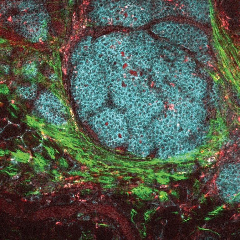
Drug-loaded nanoparticles target and kill breast cancer cells.© J. SZULCZEWSKI, D. INMAN, K. ELICEIRI, P. KEELY \ NCI \ CARBONE CANCER CENTER AT THE UNIV. OF WISCONSIN
Chemotherapeutics have markedly increased cancer survival rates. However, they cause serious side-effects due to their inability to differentiate between cancerous and healthy cells. Research aiming to reduce these adverse effects is important to ensure treatment compliance and improve patients’ quality of life.
Kheireddine El-Boubbou has been designing nanoparticles that combine diagnostic and therapeutic capabilities, also known as theranostics, for more than ten years. In a recent study published in Bioconjugate Chemistry, El-Boubbou and his team at King Saud bin Abdulaziz University for Health Sciences, King Abdullah International Medical Research Center and Saudi Arabia’s Ministry of National Guard—Health Affairs describe how novel magnetic and fluorescent nanoparticles are able to deliver an anti-cancerous drug to breast cancer cells with high selectivity.
Magnetic metal oxide nanoparticles have demonstrated considerable promise for disease diagnosis, as magnetic resonance contrast agents, and as drug delivery agents, but mass production of such particles for clinical applications has been particularly challenging.
Researchers led by El-Boubbou have developed a unique approach for producing magnetic metal oxide nanoparticles easily, cheaply and safely. Their ‘Ko-precipitation Hydrolytic Basic (KHB)’ methodology allows the synthesis of particles two to six nanometres in size from affordable, non-toxic and readily available starting materials at far lower temperatures (80 °C) than existing thermal techniques (200–300 °C). With the appropriate coating, the generated particles can be used for widespread biomedical applications, such as disease diagnosis and to aid targeted drug delivery, explains El-Boubbou.
When the researchers loaded their nanoparticles with the chemotherapy drug doxorubicin and evaluated the effects on two types of cancerous breast cells and on healthy cells the results surpassed expectations. Not only were the drug-loaded particles more effective at killing the cancerous cells than the free doxorubicin, but, more importantly, their effect was greater on the cancerous cells than on the non-cancerous cells.
Thanks to the particles’ magnetic and fluorescent properties, the authors were able to electronically image the drug-loaded nanoparticles and demonstrate cell uptake, even in tumour tissue derived from patients with breast cancer. Unlike free doxorubicin, which is internalised by passive diffusion through the cell membrane, the drug-loaded nanoparticles trigger endocytosis, a process whereby the cell actively engulfs them.
The ability of these particles to selectively deliver the chemotherapy drug to cancerous cells is somewhat surprising given that they have not been designed against a molecular target on these cells. In El-Boubbou’s own words “these results suggest that even with a passive-targeted, well-designed delivery system, enhanced toxic responses [in cancerous cells] can be attained,” offering huge potential as selective anti-cancer drug carriers.


